Review
. 2016 Feb;1366(1):20-39.
doi: 10.1111/nyas.12794. Epub 2015 Jun 10.

Neuromyelitis optica and the evolving spectrum of autoimmune aquaporin-4 channelopathies: a decade later

Affiliations
Review

Neuromyelitis optica and the evolving spectrum of autoimmune aquaporin-4 channelopathies: a decade later

Sean J Pittock et al. Ann N Y Acad Sci. 2016 Feb.

Abstract

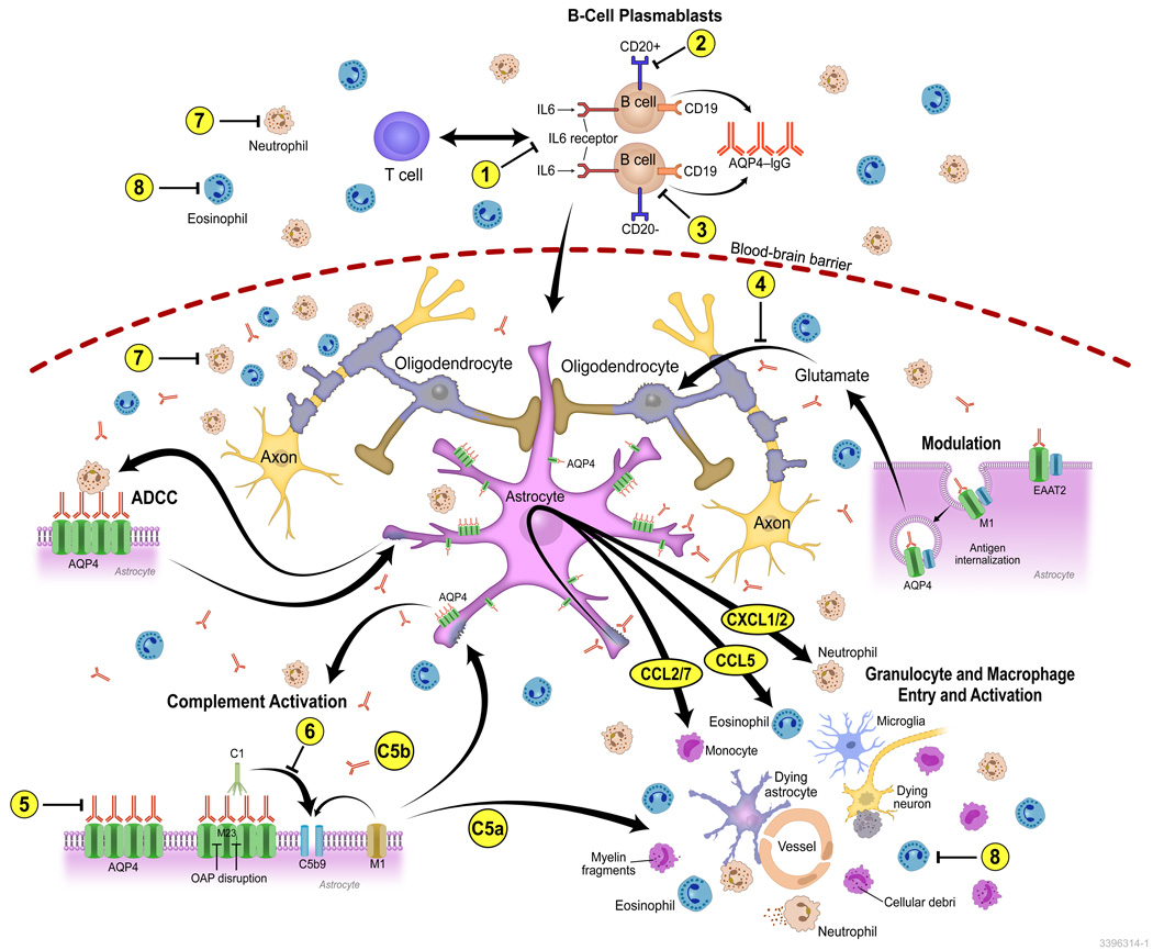
The discovery of AQP4-IgG (a pathogenic antibody that targets the astrocytic water channel aquaporin-4), as the first sensitive and specific biomarker for any inflammatory central nervous system demyelinating disease (IDD), has shifted emphasis from the oligodendrocyte and myelin to the astrocyte as a central immunopathogenic player. Neuromyelitis optica (NMO) spectrum disorders (SDs) represent an evolving spectrum of IDDs extending beyond the optic nerves and spinal cord to include the brain (especially in children) and, rarely, muscle. NMOSD typical brain lesions are located in areas that highly express the target antigen, AQP4, including the circumventricular organs (accounting for intractable nausea and vomiting) and the diencephalon (accounting for sleep disorders, endocrinopathies, and syndrome of inappropriate antidiuresis). Magnetic resonance imaging brain abnormalities fulfill Barkoff criteria for multiple sclerosis in up to 10% of patients. As the spectrum broadens, the importance of highly specific assays that detect pathogenic AQP4-IgG targeting extracellular epitopes of AQP4 cannot be overemphasized. The rapid evolution of our understanding of the immunobiology of AQP4 autoimmunity necessitates continuing revision of NMOSD diagnostic criteria. Here, we describe scientific advances that have occurred since the discovery of NMO-IgG in 2004 and review novel targeted immunotherapies. We also suggest that NMOSDs should now be considered under the umbrella term autoimmune aquaporin-4 channelopathy.

Keywords: aquaporin-4; multiple sclerosis; myelitis; neuromyelitis optica; optic neuritis.


Figures

Figure 1
Figure 1
Autoimmune aquaporin-4 (AQP4) channelopathy. Schematic for proposed diagnostic criteria incorporating the expanding clinical phenotypes of neuromyelitis optica spectrum disorders (NMOSD). This schema is based on the proposal that the core diagnostic criteria for any NMOSD requires the presence of the AQP4-immunoglobulin (IgG; red) and assumes no false positivity. Seropositivity for AQP4-IgG must be interpreted within the clinical context. The absence of the biomarker (yellow outer circle) could indicate an alternative diagnosis, such as multiple sclerosis, another demyelinating disease, or an indeterminate disorder. Some patients may have autoimmune MOG oligodendrogliopathy. Each neurological manifestation is represented by a circle. The area of the circle overlapping with the red AQP4-IgG+ circle represents an approximation of the proportion of patients with that neurological manifestation considered NMOSD (e.g., < 5% of patients with single-episode optic neuritis; 5–25% of patients with recurrent optic neuritis; 40% of patients with single-episode longitudinally extensive transverse myelitis (LETM); 70–90% of patients with recurrent LETM). The area of the circle outside (yellow) the red AQP4-IgG+ circle does not fulfill criteria for NMOSD. The arrows signify that neurological manifestations commonly coexist; for example, patients might present with or have a history of more than one neurological manifestation, such as optic neuritis and intractable vomiting, syndrome of inappropriate antidiuresis and LETM, LETM and brain stem disorder, and LETM and posterior reversible encephalopathy syndrome. Only rarely is myositis encountered. Adapted from Ref. 11 with permission from Wiley.
Figure 2
Figure 2
Mechanisms of NMO pathogenesis and pharmacologic targets. NMO lesions have a complex spectrum of characteristics indicating multiple pathogenic mechanisms. In NMOSD, the immunizing event is not known. AQP4-IgG produced by plasma cells and plasmablasts penetrates the CNS through endothelial transcytosis or at areas of relative blood–brain barrier permeability or injury (some may be produced within the CNS). Binding of NMO IgG to AQP4 on the surface of astrocytes induces an inflammatory reactive stress response that results in transcriptional and translational events within the astrocyte that promote the primary recruitment of granulocytes to the CNS. In vitro studies have demonstrated upregulation of adhesion factors such as ICAM-1 and VCAM-1, the initiation of oxidative stress response pathways involving SOD2 and ceruloplasmin, and the upregulation of inflammasome components such as caspase-1 and various proteasome subunits, inducing a massive cytokine and chemokine response (e.g., CXCL1/2 targeting neutrophils; CCL2 and CCL7 targeting monocytes, and CCL5 targeting eosinophils). Antibody-dependent astrocyte damage involving complement-dependent cytotoxicity and antibody-dependent cellular cytotoxicity (ADCC) mechanisms amplify inflammation and contribute to additional oligodendrocyte injury, demyelination, and neuronal loss. The numbers (1–8) indicate some pharmacological targets in NMO including approved drugs under evaluation for repurposing, drugs in preclinical development, and drugs at early, proof-of-concept stage: (1) IL-6 inhibitors: tocilizumab (Actemra, Toactemra; Genentech); SA237 (Chugai): phase III strial due to start enrolling patients soon; (2) CD20: rituximab; (3) CD19: MEDI-551 (Medimmune LLC), phase III trial enrolling patients; (4) glutamate antagonists; (5) AQP4-IgG blocking and inactivation strategies: high-affinity, nonpathogenic anti-AQP4 antibody (aquaporumab) competes with pathogenic AQP4-IgG for AQP4 binding; (6) complement activation pathways and complement drug targets: eculizumab (Soliris) inhibits C5 and is now in phase III trial; C1inh (Cinryse) inhibits C1, phase I completed; (7) anti-neutrophil: sivelestat (Elaspol), phase I and II recruiting patients; and (8) anti-eosinophil: antihistamines, cetirizine, and ketotifen reduce cytotoxicity mediated by AQP4-IgG and eosinophils in in vitro models. Eosinophil-stabilizing agents are currently being studied in small numbers of patients with NMO.
Figure 3
Figure 3
Molecular-based approach to classification of inflammatory demyelinating diseases (IDDs). Schematic for proposed classification of CNS IDDs according to the molecular target. The circles illustrate the clinical phenotypic overlap for IDDs where the molecular target is known (AQP4 in red and MOG in green) or yet to be discovered (orange circles indicate hypothetical clinical phenotypic overlap and question marks indicate clinical symptoms or signs to be determined). This schema recognizes that clinical phenotype may be shared (for example optic neuritis or transverse myelitis) though the immunopathogenic mechanisms may differ (astrocytopathy in AQP4 autoimmunity and oligodendrogliopathy in MOG autoimmunity). Recognition of such immunopathogenic differences will likely be important as more individualized and mechanistically targeted therapies become available.

References

    1. Devic E. Myélite aiguë dorso-lombaire avec névrite optique. - Autopsie. Congres francais de medecine. 1895:434–439.
    1. Gault F. De la neuromyelite optique aigue. These: Faculte de Medecine et de Pharmacie. 1894
    1. Jarius S, Wildemann B. The history of neuromyelitis optica. J Neuroinflammation. 2013;10:8. - PMC - PubMed
    1. Jarius S, Wildemann B. An early case of neuromyelitis optica: on a forgotten report by Jacob Lockhart Clarke, FRS. Mult Scler. 2011;17(11):1384–1386. - PubMed
    1. Jarius S, Wildemann B. The case of the Marquis de Causan (1804): an early account of visual loss associated with spinal cord inflammation. J Neurol. 2012;259(7):1354–1357. - PubMed

Publication types

MeSH terms