Review
. 2015 Oct;282(19):3693-721.
doi: 10.1111/febs.13342. Epub 2015 Jul 4.

Proteolytic cleavage, trafficking, and functions of nuclear receptor tyrosine kinases

Affiliations
Review

Proteolytic cleavage, trafficking, and functions of nuclear receptor tyrosine kinases

Mei-Kuang Chen et al. FEBS J. 2015 Oct.

Abstract

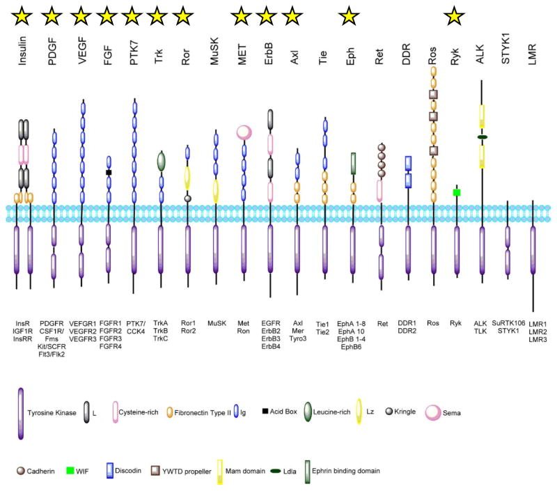
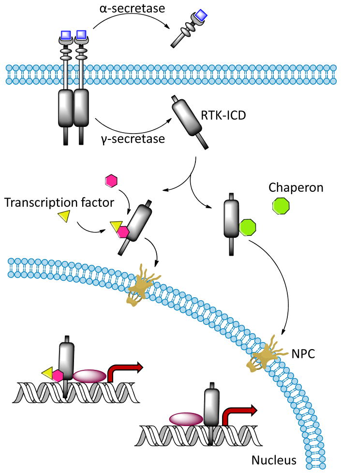
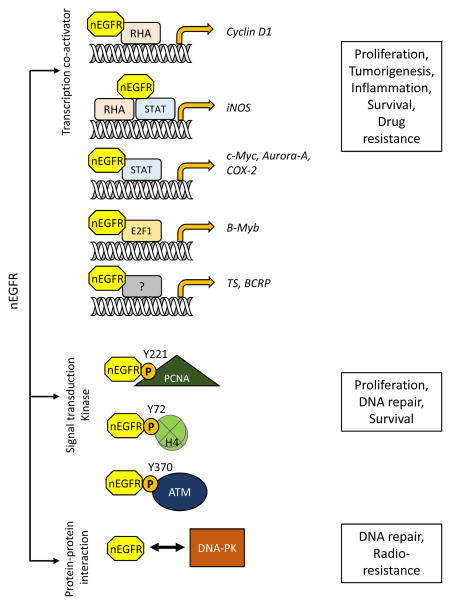
Intracellular localization has been reported for over three-quarters of receptor tyrosine kinase (RTK) families in response to environmental stimuli. Internalized RTK may bind to non-canonical substrates and affect various cellular processes. Many of the intracellular RTKs exist as fragmented forms that are generated by γ-secretase cleavage of the full-length receptor, shedding, alternative splicing, or alternative translation initiation. Soluble RTK fragments are stabilized and intracellularly transported into subcellular compartments, such as the nucleus, by binding to chaperone or transcription factors, while membrane-bound RTKs (full-length or truncated) are transported from the plasma membrane to the ER through the well-established Rab- or clathrin adaptor protein-coated vesicle retrograde trafficking pathways. Subsequent nuclear transport of membrane-bound RTK may occur via two pathways, INFS or INTERNET, with the former characterized by release of receptors from the ER into the cytosol and the latter characterized by release of membrane-bound receptor from the ER into the nucleoplasm through the inner nuclear membrane. Although most non-canonical intracellular RTK signaling is related to transcriptional regulation, there may be other functions that have yet to be discovered. In this review, we summarize the proteolytic processing, intracellular trafficking and nuclear functions of RTKs, and discuss how they promote cancer progression, and their clinical implications.

Keywords: INTERNET; MRIN; RTK cleavage; intracellular trafficking; non-canonical RTK functions; nuclear receptor tyrosine kinases; nuclear translocation; proteolytic cleavage; receptor tyrosine kinase; retrograde trafficking.


Figures

Figure 1
Figure 1. Receptor tyrosine kinase subfamilies
Among receptor tyrosine kinases, 20 subfamilies are identified in human genome. Those that contain receptor reported as MRIN are marked by a yellow star. The subfamilies are illustrated here with their extracellular domain structures marked accordingly in the key. The intracellular tyrosine kinase domain is shown as purple rectangle. The family members are noted below each subfamily. The lipid bilayer represents the plasma membrane, and the schematic here does not reflect the actual scale. (This figure is modified from the work of Lemmon and Schlessinger [3]).
Figure 2
Figure 2. Canonical RTK signaling cascade
The canonical RTK signaling begins with (1) the receptor binding with its ligand. (2) The receptor then undergoes oligomerization and trans auto-phosphorylation. (3) The phosphor-tyrosine residues serve as docking sites for the secondary messenger proteins containing either SH2 and/or PTB domain (green) that are subjected to phosphorylation by the RTK. (4) The secondary messenger proteins recruit and activate downstream proteins (burgundy) which serve as envoys delivering signals into the nucleus to regulate gene transcription.
Figure 3
Figure 3. Protease-dependent RTK-ICD formation and intracellular domain trafficking
After activation, the RTKs are subjected to α-secretase mediated shedding to free the ectodomain before intracellular domain (ICD) is released into cytoplasm by γ-secretase cleavage. The ICDs are stabilized and guided across the cytoplasm and into nucleus through nuclear pore complex (NPC) by binding to transcription factors or chaperons. ICDs have been reported to act as transcriptional coactivators.
Figure 4
Figure 4. Endosomal vesicle trafficking of internalized RTK
RTKs are internalized through either clathrin-dependent or clathrin-independent pathways. In clathrin-dependent endocytosis mechanism, the internalized membrane vesicle is coated with clathrin (green). Meanwhile, caveolin-mediated endocytosis, which is the main clathrin-independent RTK endocytic mechanism, is initiated at the membrane region that contain caveolin-rich lipid raft (purple). The endocytic vesicles from both pathways are sent to early endosome for sorting. Based on the component of coating proteins, the vesicles are then transported to different endosomal components, including recycle endosome, late endosome and trans Golgi network. Several important coating proteins that direct vesicle transport are shown, including Rab proteins, clathrin-dependent adaptor proteins (AP), retromer, syntaxin 6 (Syn 6), and Golgi-associated, gamma adaptin ear containing, ARF binding protein (GGA).
Figure 5
Figure 5. Retrograde and nuclear transport mechanisms of membrane-bound RTKs
RTKs are transported from Golgi to ER in COPI-coated vesicles via the retrograde pathway. The ER-to-nuclear transport of RTKs is mediated by two pathways, INFS and INTERNET. Via INFS pathway, the RTK is pumped through the Sec61 complex into the cytosol where it binds to cytosolic importin complex and transported into nucleus by the importin-NPC interaction. Via the INTERNET pathway, the RTK is trafficked along ER, translocated from the ONM to INM through the NPC by binding to ER-associated importin, and released from the INM by the Sec61 complex into the nucleoplasm. Nuclear localized RTK interacts with transcription factors and functions as transcriptional regulator.
Figure 6
Figure 6. Functions of nuclear EGFR
Nuclear EGFR (nEGFR) has different roles in regulating cellular fate. (1) nEGFR can function as a transcription co-activator by interacting with different transcription factors, such as RNA helicase A (RHA), STAT, and E2F1, to promote target gene transcription. Elevated levels of these genes result in cell proliferation, tumorigenesis, and inflammation and are also correlated with drug resistance. (2) nEGFR phosphorylates its substrates, such as PCNA, histone H4, and ATM, to promote cell proliferation and DNA repair. (3) nEGFR also interacts with DNA-PK to increase DNA repair, leading to the development of radioresistance.

References

    1. Keefe DM, Bateman EH. Tumor control versus adverse events with targeted anticancer therapies. Nat Rev Clin Oncol. 2012;9:98–109. - PubMed
    1. Sundaram MV. Canonical RTK-Ras-ERK signaling and related alternative pathways. WormBook. 2013:1–38. - PMC - PubMed
    1. Lemmon MA, Schlessinger J. Cell signaling by receptor tyrosine kinases. Cell. 2010;141:1117–34. - PMC - PubMed
    1. Schlessinger J. Receptor tyrosine kinases: legacy of the first two decades. Cold Spring Harb Perspect Biol. 2014;6 - PMC - PubMed
    1. Templeton AJ, Diez-Gonzalez L, Ace O, Vera-Badillo F, Seruga B, Jordan J, Amir E, Pandiella A, Ocana A. Prognostic relevance of receptor tyrosine kinase expression in breast cancer: A meta-analysis. Cancer Treat Rev 2014 - PubMed

Publication types

MeSH terms

Substances

LinkOut - more resources