. 2015 Sep 2;11(33):4173-82.
doi: 10.1002/smll.201500027. Epub 2015 Jun 11.

The Sequence-Specific Cellular Uptake of Spherical Nucleic Acid Nanoparticle Conjugates

Affiliations

The Sequence-Specific Cellular Uptake of Spherical Nucleic Acid Nanoparticle Conjugates

Suguna P Narayan et al. Small. .

Abstract

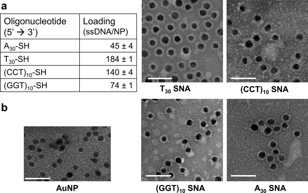
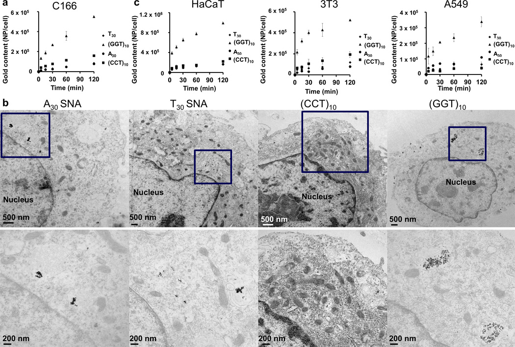
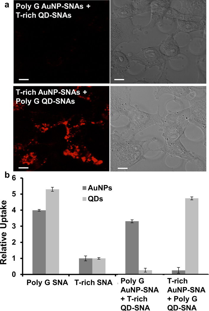
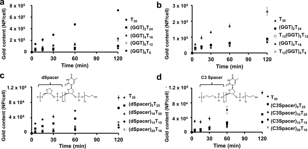
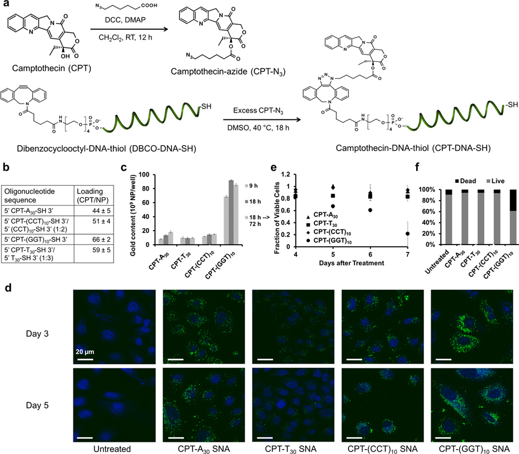
The sequence-dependent cellular uptake of spherical nucleic acid nanoparticle conjugates (SNAs) is investigated. This process occurs by interaction with class A scavenger receptors (SR-A) and caveolae-mediated endocytosis. It is known that linear poly(guanine) (poly G) is a natural ligand for SR-A, and it has been proposed that interaction of poly G with SR-A is dependent on the formation of G-quadruplexes. Since G-rich oligonucleotides are known to interact strongly with SR-A, it is hypothesized that SNAs with higher G contents would be able to enter cells in larger amounts than SNAs composed of other nucleotides, and as such, cellular internalization of SNAs is measured as a function of constituent oligonucleotide sequence. Indeed, SNAs with enriched G content show the highest cellular uptake. Using this hypothesis, a small molecule (camptothecin) is chemically conjugated with SNAs to create drug-SNA conjugates and it is observed that poly G SNAs deliver the most camptothecin to cells and have the highest cytotoxicity in cancer cells. Our data elucidate important design considerations for enhancing the intracellular delivery of spherical nucleic acids.

Keywords: cellular uptake; guanine; nanoparticles; sequence-specific; spherical nucleic acids.


Figures

Figure 1
Figure 1
Characterization of SNAs. a) The table lists the loading of oligonucleotides on a 10 nm gold nanoparticle using a fluorescence-based assay. Poly T SNAs contain the highest loading among all nucleobase types, whereas poly A SNAs have the lowest. b) Staining of SNAs by uranyl acetate clearly delineates the DNA oligonucleotide shell (white) around the gold nanoparticle core (black) by TEM imaging. The thickness of the shell correlates with the oligonucleotide loading data obtained from the fluorescence-based assay. Scale bar = 50 nm.
Figure 2
Figure 2
Cellular uptake of SNAs. a) Poly G SNAs show the highest association with C166 cells, 4–10 times higher than SNAs composed of other nucleobase types (p < 0.001). b) By TEM imaging, poly G SNAs exhibit the highest accumulation inside C166 cells, as evidenced by their widespread distribution throughout the cytosol as large clusters (>100 per clusters). By contrast, SNAs composed of other nucleobase types either accumulate in more confined regions of the cytosol or appear in clusters that contain fewer particles (<20 particles per cluster). The bottom row features enlarged images of the boxed regions of the top row. c) Poly G SNAs also demonstrate the highest association with three other cell lines beside C166, including, in descending order of expression level for SR-A, HaCaT (immortal human keratinocyte), 3T3 (mouse fibroblast), and A549 (human lung epithelial adenocarcinoma). For all cell types, poly G SNAs exhibit 3–5 times higher association with cells than SNAs of other nucleobase types (p < 0.001). Association of poly G SNAs with cells positively correlates with the expression level of SR-A for the same cell types. Error bars denote the standard deviation from triplicate measurements.
Figure 3
Figure 3
Dependence of uptake on poly G shell. a) By confocal microscopy, poly G QD-SNAs (red) show higher accumulation in C166 cells compared to T-rich QD-SNAs. Scale bar = 10 µm. b) ICP-MS analysis of the gold and cadmium content in C166 cells co-treated with T-rich AuNP-SNAs and poly G QD-SNAs as well as co-treated with T-rich QD-SNAs and poly G AuNP-SNAs shows that poly G AuNP-SNAs preferentially enter cells compared to T-rich QD-SNAs and poly G QD-SNAs preferentially enter cells compared to T-rich AuNP-SNAs (p < 0.001). Error bars denote the standard deviation from three independent experiments.
Figure 4
Figure 4
Length of oligonucleotide strand affecting cellular uptake of SNAs. a) Increased guanine (G) content at the 5’ end of constituent oligonucleotides increases cellular association of SNAs with C166 cells (p < 0.001). A minimum of four GGT repeating units is necessary to enhance cellular association of SNAs when compared to poly T (T30) SNAs. b) Burial of the GGT repeating units in the middle of the constituent oligonucleotides negates the enhancement in cellular association (p < 0.001). c) Increasing dSpacer units (which do not have a nucleobase) at the 5’ end of constituent DNA oligonucleotides reduces cellular association of SNAs up to 75% (p < 0.001). d) Increasing C3 Spacer units (which have neither a nucleobase nor a ribose) at the 5’ end of constituent DNA oligonucleotides reduces cellular association of SNAs up to 75% (p < 0.001). Error bars denote the standard deviation from triplicate measurements.
Figure 5
Figure 5
Delivery of camptothecin molecules using CPT-SNAs. a) The -OH group of the camptothecin molecule (CPT) is modified by a short bifunctional linker to form camptothecin azide (CPT-N3) by literature precedent. CPT-N3 is then coupled to dibenzocyclooctyl-DNA-thiol (DBCO-DNA-SH) by copper-free click chemistry to form camptothecin-DNA-thiol (CPT-DNA-SH). DCC = N’ N’-dicyclohexocarbodiimide, DMAP = 4-dimethylaminopyridine, CH2Cl-2 = dichloromethane, DMSO = dimenthyl sulfoxide. b) Measurements based on the fluorescence emission of CPT at 440 nm revealed that CPT-SNAs of all four nucleobase types contain 55 ± 15 CPT molecules per particle. c) By ICP-MS analysis of the gold content of the A549 cells treated with CPT-SNAs, CPT-poly G SNAs can enter cells in highest quantities among all nucleobase types tested (p < 0.001). The CPT-SNAs (at least the AuNP core) do not seem to leave the cells after the treatment. Error bars denote the standard deviation from triplicate measurements. d) By confocal imaging, CPT-poly G SNAs can deliver CPT molecules (green) into A549 cells in highest quantities among CPT-SNAs of all nucleobase types tested. Blue = nucleus. Scale bar = 20 µm. By the MTT assay (e) and flow cytometry analysis supported by propidium iodide staining (f), CPT-poly G SNAs are also most cytotoxic among CPT-SNAs of all nucleobase types tested after 6 days (p < 0.01). Error bars denote the standard deviation from four measurements.

References

    1. Cutler JI, Auyeung E, Mirkin CA. J Am Chem Soc. 2012;134:1376–1391. - PubMed
    1. Rosi NL, Giljohann DA, Thaxton CS, Lytton-Jean AK, Han MS, Mirkin CA. Science. 2006;312:1027–1030. - PubMed
    1. Seferos DS, Giljohann DA, Hill HD, Prigodich AE, Mirkin CA. J Am Chem Soc. 2007;129:15477–15479. - PMC - PubMed
    1. Giljohann DA, Seferos DS, Prigodich AE, Patel PC, Mirkin CA. J Am Chem Soc. 2009;131:2072–2073. - PMC - PubMed
    1. Massich MD, Giljohann DA, Seferos DS, Ludlow LE, Horvath CM, Mirkin CA. Mol Pharm. 2009;6:1934–1940. - PMC - PubMed

Publication types