. 2015 Nov;67(11):2990-3003.
doi: 10.1002/art.39247.

Release of neutrophil extracellular traps by neutrophils stimulated with antiphospholipid antibodies: a newly identified mechanism of thrombosis in the antiphospholipid syndrome

Affiliations

Release of neutrophil extracellular traps by neutrophils stimulated with antiphospholipid antibodies: a newly identified mechanism of thrombosis in the antiphospholipid syndrome

Srilakshmi Yalavarthi et al. Arthritis Rheumatol. 2015 Nov.

Abstract

Objective: Antiphospholipid antibodies (aPL), especially those targeting β2 -glycoprotein I (β2 GPI), are well known to activate endothelial cells, monocytes, and platelets, with prothrombotic implications. In contrast, the interaction of aPL with neutrophils has not been extensively studied. Neutrophil extracellular traps (NETs) have recently been recognized as an important activator of the coagulation cascade, as well as an integral component of arterial and venous thrombi. This study was undertaken to determine whether aPL activate neutrophils to release NETs, thereby predisposing to the arterial and venous thrombosis inherent in the antiphospholipid syndrome (APS).

Methods: Neutrophils, sera, and plasma were prepared from patients with primary APS (n = 52) or from healthy volunteers and characterized. No patient had concomitant systemic lupus erythematosus.

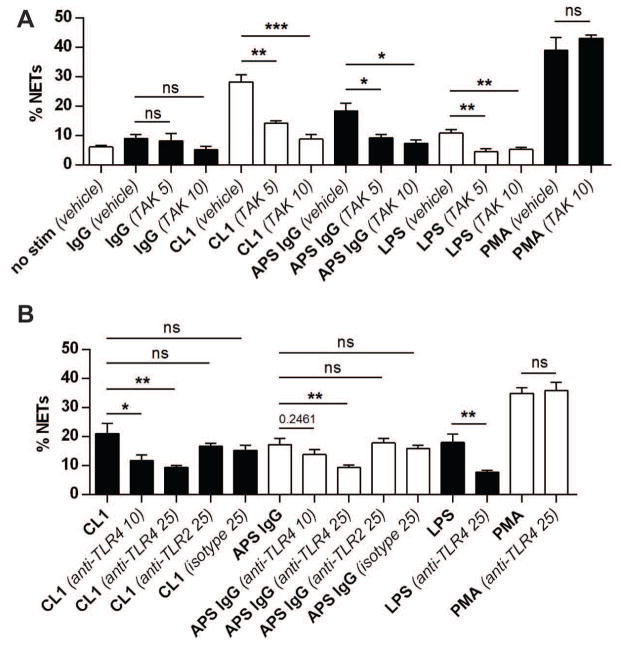
Results: Sera and plasma from patients with primary APS had elevated levels of both cell-free DNA and NETs, as compared to healthy volunteers. Freshly isolated neutrophils from patients with APS were predisposed to high levels of spontaneous NET release. Further, APS patient sera, as well as IgG purified from APS patients, stimulated NET release from control neutrophils. Human aPL monoclonal antibodies, especially those targeting β2 GPI, also enhanced NET release. The induction of APS NETs was abrogated with inhibitors of reactive oxygen species formation and Toll-like receptor 4 signaling. Highlighting the potential clinical relevance of these findings, APS NETs promoted thrombin generation.

Conclusion: Our findings indicate that NET release warrants further investigation as a novel therapeutic target in APS.


Conflict of interest statement

CONFLICT OF INTEREST DISCLOSURES

The authors have no competing interests or conflicts to disclose.

Figures

Figure 1
Figure 1. Cell-free DNA and NETs are increased in the circulation of patients with primary antiphospholipid syndrome (APS)
A, Cell-free DNA was measured in the plasma of APS patients (n=26) or healthy controls. B, Circulating NETs were measured in the same plasma by MPO-DNA ELISA. C, Cell-free DNA was measured in sera of APS patients (n=52) or healthy controls. D, Correlation between cell-free DNA and circulating NETs (MPO-DNA complexes) in APS sera. *p<0.05 and **p<0.01. In addition to the individual data points, mean and standard deviation are also presented in panels A–C.
Figure 2
Figure 2. Primary APS neutrophils demonstrate enhanced NET release
A, Freshly-isolated neutrophils from healthy controls or APS patients (n=18) were seeded onto coverslips and incubated in serum-free media for 2 hours. NET release was scored by immunofluorescence microscopy. B, Representative immunofluorescence microscopy of control (top) and APS (bottom) neutrophils as presented in panel A. Blue=DNA and green=neutrophil elastase. Scale bars=25 microns. C, Control neutrophils were treated with 10% sera from heterologous healthy controls or APS patients for 2 hours. NET release was scored by immunofluorescence microscopy. *p<0.05, ***p<0.001, and ****p<0.0001; ns=not significant. In addition to individual data points, mean and standard deviation are also presented.
Figure 3
Figure 3. Anti-β2GPI IgG stimulates neutrophils to release NETs
A, Five APS IgG samples were pooled, and then depleted of anti-β2GPI IgG using purified β2GPI protein. Control neutrophils were stimulated with IgG (10 ug/ml) as indicated for 3 hours. NET release was scored by immunofluorescence microscopy. B, Control neutrophils were treated with purified monoclonal antiphospholipid Abs (aPL; 10 μg/ml) for 3 hours in the presence or absence of 10% autologous serum. aPL IS4, CL1, and CL24 are known to bind β2GPI, while IS1 and IS2 do not. P values were determined by comparing data sets to control IgG (−/+ serum as appropriate). C, Control neutrophils were stimulated with control IgG, CL1, or 20 nM PMA. After 2 hours, PicoGreen was added to the culture, and fluorescence intensity (corresponding to extracellular DNA) was measured. Normalization was to cells permeablized with 0.1% Triton. In panels A–C, bars represent the mean and SEM of at least five independent experiments; *p<0.05, **p<0.01, ***p<0.001, ****p<0.0001. D, Representative live cells stained with PicoGreen as in panel C. In the CL1 sample, expanded cell remnants (dashed lines) are surrounded by a halo of DNA (green). Scale bars=25 microns.
Figure 4
Figure 4. β2GPI is detectable on the neutrophil surface
A, Neutrophils and peripheral blood mononuclear cells (PBMCs) were isolated from healthy controls. Total protein extracts were prepared by detergent lysis. Western blotting was to β2GPI, annexin A2, and β-actin. Quantification was by densitometry and is expressed in arbitrary units. Bars represent mean and SEM. N=6, including 3 samples for each group not pictured here; western blotting was repeated twice with similar results. ***p<0.001. B, Neutrophils were isolated from healthy controls and allowed to adhere to coverslips. Cells were then immediately fixed with paraformaldehyde, and in some cases permeabilized with detergent (0.1% Triton). Representative images are shown with β2GPI and neutrophil elastase stained green and DNA stained blue. Scale bars=25 microns. C, Neutrophils and monocytes were identified by forward/side-scatter, and additionally confirmed to be CD10-positive and CD14-positive, respectively. The percentage of β2GPI-positive cells was then determined. Bars represent mean and SEM. N=6 healthy controls for each group; **p<0.01. To the right is a representative neutrophil histogram, demonstrating that the majority of CD10-positive cells are also positive for β2GPI.
Figure 5
Figure 5. aPL-mediated NET release can be blocked by inhibition of TLR4, but not TLR2
A, Control neutrophils were stimulated with control human IgG, an anti-β2GPI monoclonal (CL1), or pooled IgG from 5 APS patients (all 10 μg/ml) for 3 hours. Control treatments were with LPS (100 ng/ml) and PMA (20 nM). Some samples were pretreated with TAK-242, a TLR4 inhibitor, at 5 or 10 μM. NET release was scored by immunofluorescence microscopy. B, Control neutrophils were stimulated as indicated (concentrations as in panel A) for 3 hours. Some samples were pretreated with anti-TLR4, anti-TLR2, or isotype (10 and 25 μM). NET release was scored by immunofluorescence microscopy. For all panels, bars represent mean and SEM; n=5 independent experiments per condition. *p<0.05, **p<0.01, ***p<0.001, and ns=not significant.
Figure 6
Figure 6. Purified aPL, as well as APS patient plasma, stimulate thrombin generation in neutrophil- and DNA-dependent fashion
A, Representative thrombin (IIa) generation plot demonstrating enhanced generation when neutrophils are exposed to platelet-poor plasma (from a healthy control) supplemented with aPL CL1 (10 μg/ml). The effect is not seen with plasma alone or plasma supplemented with control IgG. The effect is disrupted by DNase treatment. B, Representative data demonstrating that control plasma supplemented with anti-β2GPI monoclonals promotes neutrophil-mediated thrombin generation. Delta (Δ) values were calculated relative to the baseline condition (in this case, plasma supplement with IgG, but not neutrophils). These data are representative of 3 experiments, all with similar results. C, Control plasma was separately supplemented with total IgG (10 μg/ml) isolated from 5 healthy controls or 5 primary APS patients with anti-β2GPI IgG positivity. Plasma was then mixed with control neutrophils alone or neutrophils + DNase, and thrombin generation was determined. D, Plasma from healthy controls or primary APS patients was mixed with control neutrophils alone or neutrophils + DNase, and thrombin generation was determined. In panels C and D, mean and standard deviation are presented; *p<0.05 and **p<0.01. Each data point is the average of 3 independent assays.

Comment in

References

    1. Bertolaccini ML, Amengual O, Andreoli L, Atsumi T, Chighizola CB, Forastiero R, et al. Autoimmunity reviews; 14th International Congress on Antiphospholipid Antibodies Task Force. Report on antiphospholipid syndrome laboratory diagnostics and trends; 2014. - PubMed
    1. Hughes GR. Hughes’ syndrome: the antiphospholipid syndrome. A historical view. Lupus. 1998;7 (Suppl 2):S1–4. - PubMed
    1. Erkan D, Aguiar CL, Andrade D, Cohen H, Cuadrado MJ, Danowski A, et al. 14th International Congress on Antiphospholipid Antibodies: task force report on antiphospholipid syndrome treatment trends. Autoimmunity reviews. 2014;13(6):685–96. - PubMed
    1. Conti F, Sorice M, Circella A, Alessandri C, Pittoni V, Caronti B, et al. Beta-2-glycoprotein I expression on monocytes is increased in anti-phospholipid antibody syndrome and correlates with tissue factor expression. Clinical and experimental immunology. 2003;132(3):509–16. - PMC - PubMed
    1. Caronti B, Calderaro C, Alessandri C, Conti F, Tinghino R, Palladini G, et al. Beta2-glycoprotein I (beta2-GPI) mRNA is expressed by several cell types involved in anti-phospholipid syndrome-related tissue damage. Clinical and experimental immunology. 1999;115(1):214–9. - PMC - PubMed

Publication types

MeSH terms