. 2015 Winter;144(1):67-82.
doi: 10.1162/DAED_a_00318.

Sleep, Memory & Brain Rhythms

Affiliations

Sleep, Memory & Brain Rhythms

Brendon O Watson et al. Daedalus. 2015 Winter.

Abstract

Sleep occupies roughly one-third of our lives, yet the scientific community is still not entirely clear on its purpose or function. Existing data point most strongly to its role in memory and homeostasis: that sleep helps maintain basic brain functioning via a homeostatic mechanism that loosens connections between overworked synapses, and that sleep helps consolidate and re-form important memories. In this review, we will summarize these theories, but also focus on substantial new information regarding the relation of electrical brain rhythms to sleep. In particular, while REM sleep may contribute to the homeostatic weakening of overactive synapses, a prominent and transient oscillatory rhythm called "sharp-wave ripple" seems to allow for consolidation of behaviorally relevant memories across many structures of the brain. We propose that a theory of sleep involving the division of labor between two states of sleep-REM and non-REM, the latter of which has an abundance of ripple electrical activity-might allow for a fusion of the two main sleep theories. This theory then postulates that sleep performs a combination of consolidation and homeostasis that promotes optimal knowledge retention as well as optimal waking brain function.


Figures
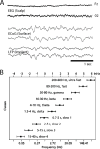
Figure 1
Figure 1. Electrical Oscillations in the Brain
(A) Recordings of brain waves occurring over approximately three seconds. Each line is a recording from one electrode with abcissa representing time and ordinate representing voltage. Top two lines are recorded from outside the skull (EEG), the middle two lines are recordings from inside the skull but on the brain surface (ECoG; electrocorticography), and the bottom two are recorded from electrodes inside the brain. (B) Illustration of the families, or “bands,” of oscillatory rhythms in the brain; each is labeled with a horizontal bar. Note that a system of rhythms is formed with a logarithmic relationship among the constituent oscillations. Source: (A) courtesy of Gregory Worrell of the Mayo Clinic and Scott Makeig of the University of California, San Diego; (B) from György Buzsáki and Andreas DraguhnNeuronal Oscillations in Cortical Networks,” Science 304 (2004): 1926–1929.
Figure 2
Figure 2. Stages of Sleep
Top panel is a graph of the depth of sleep (depth greater and arousability less toward bottom of the graph) showing a cyclic alternation between SWS and REM sleep. At bottom are example tracings for each state. Note the difference in brain wave amplitude and frequency across states. Source: György Buzsaki, Rhythms of the Brain (New York: Oxford University Press, 2006).
Figure 3
Figure 3. Schematic Replay of Waking Neuronal Activity during Sleep
At left: during waking states, experience in the environment leads to certain sequences of neuronal firing, as in this example where a rat has a number of neurons fire in a sequence corresponding to places the animal has visited. At right: fast replay of the same firing sequence of neuronal activation during a sharp wave ripple in sleep. Experiments have shown that replay after waking experience is greater than prior to waking experience. Source: adapted with permission from Gabrielle Girardeau and Michaël ZugaroHippocampal Ripples and Memory Consolidation,” Current Opinion in Neurobiology 21 (3) (2011): 452–459.
Figure 4
Figure 4. Complementary Roles for SWS and REM in Neuronal Physiology
In two types of neurons studied (pyramidal cells and inhibitory interneurons), SWS led to a slight increase of spiking activity and possibly synaptic connectivity (rising slopes), while during REM sleep, action potential generation and possibly synaptic connectivity decreased (falling slopes). Source: A. D. Grosmark, K. Mizuseki, E. Pastalkova, K. Diba, and G. Buzsakirem Sleep Reorganizes Hippocampal Excitability,” Neuron 75 (2012): 1001–1007.

References

    1. Rasch Björn, Born Jan. About Sleep’s Role in Memory. Physiological Reviews. 2013;93:681–766. - PMC - PubMed
    1. Dehaene Stanislas, Changeux Jean-Pierre. Experimental and Theoretical Approaches to Conscious Processing. Neuron. 2011;70:200–227. - PubMed
    2. Tononi G, Edelman GM. Consciousness and Complexity. Science. 1998;282:1846–1851. - PubMed
    1. Varela Francisco, Lachaux Jean-Philippe, Rodriguez Eugenio, Martinerie Jacques. The Brainweb: Phase Synchronization and Large-Scale Integration. Nature Reviews Neuroscience. 2001;2:229–239. - PubMed
    2. Engel Andreas K, Fries Pascal, Singer Wolf. Dynamic Predictions: Oscillations and Synchrony in Top-Down Processing. Nature Reviews Neuroscience. 2001;2:704–716. - PubMed
    1. Buzsáki György. Rhythms of the Brain. New York: Oxford University Press; 2006.
    1. Buzsáki György, Draguhn Andreas. Neuronal Oscillations in Cortical Networks. Science. 2004;304:1926–1929. - PubMed

LinkOut - more resources