

Case Reports
doi: 10.4103/0973-029X.157207.
Linear IgA dermatosis adult variant with oral manifestation: A rare case report
Affiliations
- PMID: 26097313
- PMCID: PMC4451675
- DOI: 10.4103/0973-029X.157207
Item in Clipboard
Case Reports
Linear IgA dermatosis adult variant with oral manifestation: A rare case report
J Oral Maxillofac Pathol.
2015 Jan-Apr.
Abstract
Linear immunoglobulin A (IgA) dermatosis (LAD) is a rare autoimmune disorder that presents as a vesiculo-bullous lesion with cutaneous manifestations, but rare oral mucosal involvement. Here we discuss a case of a vesiculobullous lesion with severe oral and ocular mucosal involvement mimicking pemphigoid with histopathological evidence of subepithelial blisters. Direct immunofluorescence (DIF) confirmed the lesion as LAD of adult variant, although with atypical clinical features.
Keywords: Cicatricial pemphigoid; linear IgA dermatosis; vesiculobullous lesion.
Conflict of interest statement
Figures

Ruptured vesicle in healing phase with scar formation in the neck
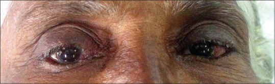
Scaring of bulbar conjunctiva on the right eye

Multiple intact bullae on hard palate with erythematous irregular ulcers

(a) Generalized desquamative gingivitis involving maxilla. (b) Generalized desquamative gingivitis involving mandible

Parakeratinized stratified squamous epithelium with subepithelial split and inflammatory infiltrate in connective tissue (H&E stain, x100)

(a) Direct immunofluorescence showing linear deposition of immunoglobulin A (IgA) at the basement membrane (Direct immunofluorescence, x 100). (b) Direct immunofluorescence showing linear deposition of immunoglobulin A (IgA) at the basement membrane (Direct immunofluorescence, x 100)

Healing of oral lesion after medication
References
-
- Tsai IC, Chu CY, Chen HJ, Wang LF, Chiu HC. Linear IgA bullous dermatosis: A clinical study of 16 cases at National Taiwan University Hospital. Dermatol Sin. 2010;28:21–6.
-
- Angiero F, Benedicenti S, Crippa R, Magistro S, Farronato D, Stefani M. Rare case of desquamative gingivitis due to linear IGA disease: Morphological and immunofluorescence features. In Vivo. 2007;21:1093–8. - PubMed
-
- Cohen LM, Skopicki DK, Harrist TJ, Clark WH., Jr . Infectious vesiculobullous and vesiculopustular diseases. In: Elder D, Elenitsas R, Jaworsky C, Johnson B, editors. Levers's Histopathology of Skin. 8th ed. LippinCott: Raven Publisher; 1997. pp. 235–7.
-
- Bickle K, Roark TR, Hsu S. Autoimmune bullous dermatoses: A review. Am Fam Physician. 2002;65:1861–70. - PubMed
Publication types
LinkOut - more resources
Full Text Sources
Miscellaneous
