Case Reports
. 2015 Jan-Apr;19(1):111.
doi: 10.4103/0973-029X.157222.

Pleomorphic adenoma of the buccal salivary gland

Affiliations
Case Reports

Pleomorphic adenoma of the buccal salivary gland

Shubhangi Khandekar et al. J Oral Maxillofac Pathol. 2015 Jan-Apr.

Abstract

Salivary gland swellings can result from tumors, an inflammatory process or cysts. It can sometimes be difficult to establish; whether pathology arises from the salivary gland itself or adjacent structures. Neoplasms of the salivary glands account for less than 1% of all tumors, 3-5% of all head and neck tumors and benign pleomorphic adenoma (PA) of minor salivary glands arising de novo is very rare. PA is the most common tumor of the salivary gland. While the majority arises from the parotid gland, only a small percentage arises from the buccal minor salivary gland. A case of PA of minor salivary glands in the buccal mucosa in a 70-year-old female is discussed. It includes review of literature, clinical features, histopathology, radiological findings and treatment of the tumor; with emphasis on diagnosis.

Keywords: Buccal minor salivary gland; chondromyxoid stroma; pleomorphic adenoma.


Conflict of interest statement

Conflict of Interest: None declared.

Figures

Figure 1
Figure 1
Extraoral photograph showing swelling and extension over right buccal region
Figure 2
Figure 2
Intraoral photograph showing no significant finding
Figure 3
Figure 3
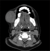
Computed tomography (CT) suggestive of heterogeneously enhancing lesion in close proximity to superficial lobe of right parotid gland without invasion of the adjacent structures
Figure 4
Figure 4
(a) Excisional biopsy of the resected tumor mass. (b) Cut sections reveals homogeneous white glistening cut potato appearance. (c) Cut sections show scattered areas of hemorrhages and necrosis
Figure 5
Figure 5
Scanner view showing lesional tissue which is encapsulated (H&E stain, x40) H&E = Hematoxylin and eosin
Figure 6
Figure 6
Scanner view showing duct like spaces with eosinophilic coagulum. (H&E stain, x40)
Figure 7
Figure 7
Low-power view showing myxochondroid areas having clear cells with eccentric nuclei (H&E stain, x100)
Figure 8
Figure 8
Low-power view showing pleomorphic and hyperchromatic cells with few mitotic figures (H&E stain, x100)
Figure 9
Figure 9
Low -power view showing the clear cells and hyalinized areas. (H&E stain, x200)
Figure 10
Figure 10
Higher-power view showing the numerous malignant pleomorphic and hyperchromatic cells with osteoid tissue and squamous metaplasia (H&E stain, x400)
Figure 11
Figure 11
Higher-power view showing spindle-shaped cells with duct-like spaces and eosinophilic coagulum with few clear cells (H&E stain, x400)
Figure 12
Figure 12
Higher-power view showing spindle-shaped cells with duct-like places filled with eosinophilic coagulum, with necrotic areas and clear cells at places (H&E stain, x400)
Figure 13
Figure 13
Higher-power view showing spindle-shaped cells with duct-like places filled with eosinophilic coagulum (H&E stain, x400)

References

    1. Greenberg M, Glick M. Burket's Oral Medicine. 11th ed. Philadelphia: Lippincott; 2008. p. 172.
    1. Rajendran R. Tumors of the Salivary Glands. In: Rajendran R, Sivapathasundaram B, editors. Shafer's Text Book of Oral Pathology. 5th ed. New Delhi: Elsevier; 2006. pp. 311–6.
    1. Carlson ER, Ord RA. 1st ed. USA: Wiley-Blackwell; 2008. Textbook and Color Atlas of Salivary Gland Pathology Diagnosis and Management; p. 228.
    1. Nanci A. 8th ed. St. Louis: Missouri Mosby an Imprint of Elsevier; 2013. Tencate's Oral histology- Development, Structure and Function; p. 276.
    1. Klijanienko J, Vielh P, Batsakis JG. Salivary Gland Tumours. 1st ed. Vol. 15. New Delhi: Karger Publishers; 2000. p. 10. - PubMed

Publication types