. 2015 Jul 1;125(7):2851-60.
doi: 10.1172/JCI79956. Epub 2015 Jun 22.

DNA methylation directs functional maturation of pancreatic β cells

DNA methylation directs functional maturation of pancreatic β cells

Sangeeta Dhawan et al. J Clin Invest. .

Abstract

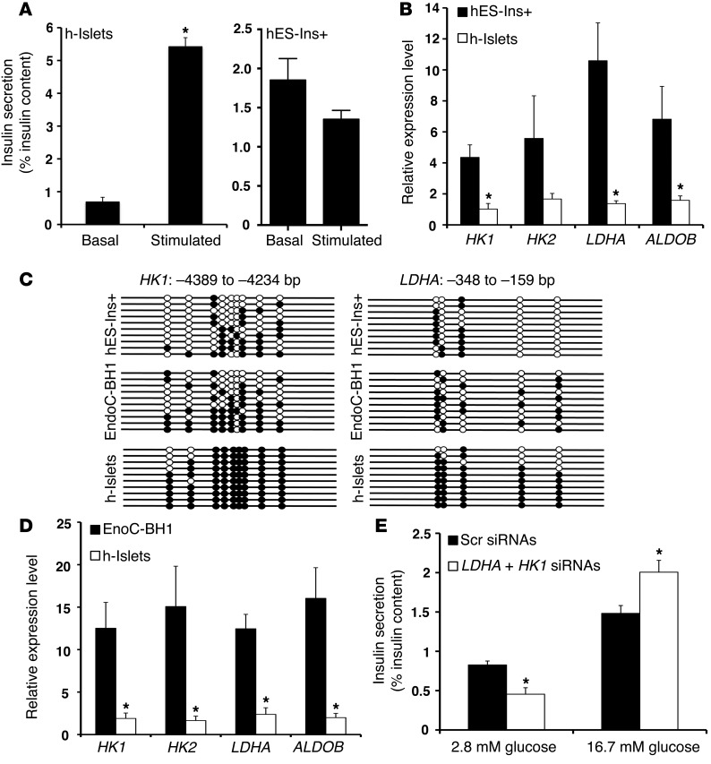
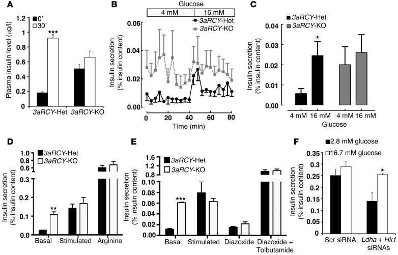
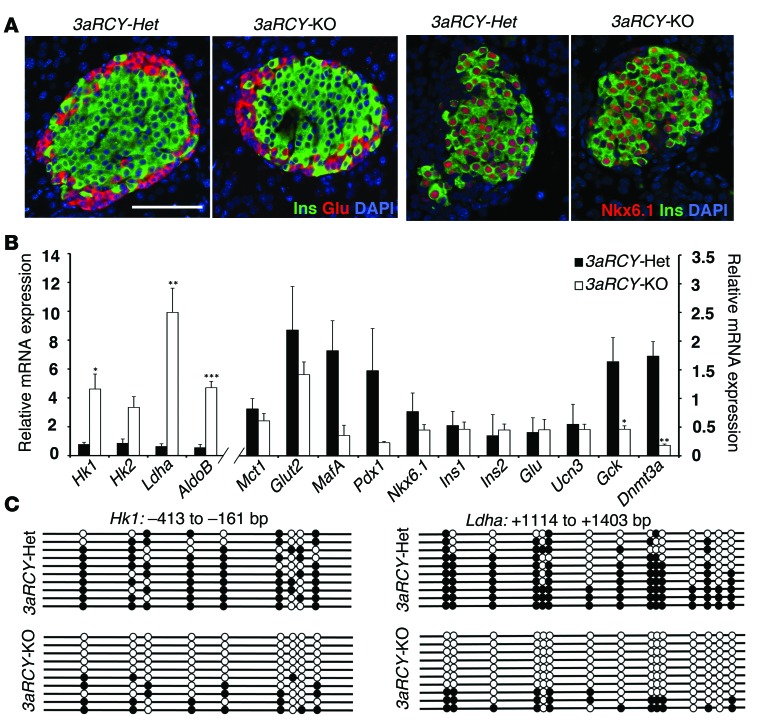
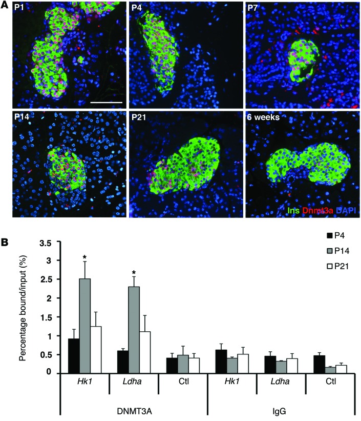
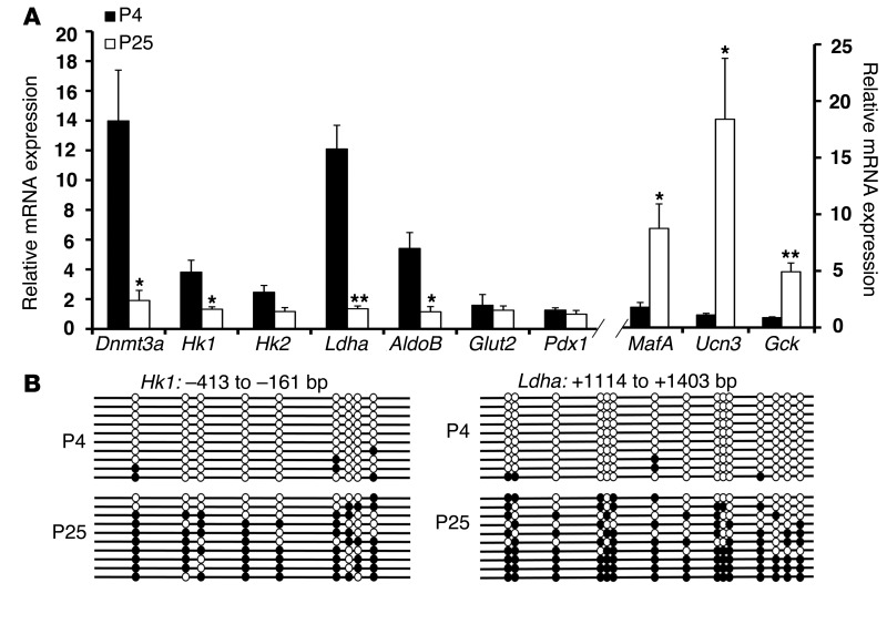
Pancreatic β cells secrete insulin in response to postprandial increases in glucose levels to prevent hyperglycemia and inhibit insulin secretion under fasting conditions to protect against hypoglycemia. β cells lack this functional capability at birth and acquire glucose-stimulated insulin secretion (GSIS) during neonatal life. Here, we have shown that during postnatal life, the de novo DNA methyltransferase DNMT3A initiates a metabolic program by repressing key genes, thereby enabling the coupling of insulin secretion to glucose levels. In a murine model, β cell-specific deletion of Dnmt3a prevented the metabolic switch, resulting in loss of GSIS. DNMT3A bound to the promoters of the genes encoding hexokinase 1 (HK1) and lactate dehydrogenase A (LDHA) - both of which regulate the metabolic switch - and knockdown of these two key DNMT3A targets restored the GSIS response in islets from animals with β cell-specific Dnmt3a deletion. Furthermore, DNA methylation-mediated repression of glucose-secretion decoupling genes to modulate GSIS was conserved in human β cells. Together, our results reveal a role for DNA methylation to direct the acquisition of pancreatic β cell function.


Figures

Figure 5
Figure 5. The metabolic programming directed by DNA methylation to regulate GSIS is conserved in human β cells.
(A) Static incubation GSIS assay, showing insulin secretion (percentage of insulin content) in differentiated insulin-positive (Ins+) human ES cells (hES-Ins+ cells; left panel), and human islets (h-islets; right panel) at 2.8 mM glucose and 16.7 mM glucose. (B) Relative mRNA expression of indicated genes in sorted GFP+ hES-Ins+ cells, compared with human islets from cadaveric donors (average donor age 43 years; n = 3). CYCLOPHILIN A was used as a housekeeping gene. The error bars represent SEM. *P < 0.05. (C) Bisulfite sequencing analysis for the HK1 and LDHA loci at indicated regions comparing sorted GFP+ hES-Ins+ cells, and EndoC-BH1 human β cell line, compared with human islets from cadaveric donors (representative clones from n = 3 samples per group). Each horizontal line with dots is an independent clone These regions are almost fully DNA methylated (filled circles) in human islets but largely hypomethylated (open circles) in hES-Ins+ cells and the EndoC-BH1 human β cell line. (D) Relative mRNA expression of indicated genes in EndoC-BH1 human β cell line, compared with human islets from cadaveric donors (average donor age 43 years). CYCLOPHILIN A was used as a housekeeping gene. n = 3 independent experiments. The error bars represent SEM. *P < 0.05. (E) Static incubation GSIS assay, showing insulin secretion (percentage of insulin content) in comparing EndoCBH1 cells treated with either a combination of siRNAs targeting HK1 and LDHA, or a scrambled (Scr) siRNA, at 2.8 mM glucose and 16.7 mM glucose. (n = 3). Error bars indicate ± SEM, *P < 0.05, Student’s t test.
Figure 4
Figure 4. β Cells lacking Dnmt3a are functionally immature.
(A) Plasma insulin levels in 6-week-old 3aRCY-KO mice and littermate-control 3aRCY-Het mice (fasted 6 hours) at 0 and 30 minutes after i.p. injection of 2 g/kg glucose (n = 3 for each genotype). (B) Insulin secretion (percentage of insulin content) in a dynamic islet perifusion assay comparing islets from 6-week-old 3aRCY-KO or control 3aRCY-Het littermates at 4 mM and 16 mM glucose. (C) Average insulin secretion (percentage of content) in the dynamic islet perifusion GSIS at 4 mM and 16 mM glucose, comparing 3aRCY-KO islets with control 3aRCY-Het islets from n = 3 mice aged 6 weeks. (D) Static incubation GSIS assay, comparing insulin secretion (percentage of insulin content) in islets from 3aRCY-KO mice with control 3aRCY-Het islets (n = 3 mice), at 2.8 mM glucose, 16.7 mM glucose, and 20 mM arginine. (E) Static incubation GSIS assay, comparing insulin secretion (percentage of insulin content) in islets from 6-week-old 3aRCY-KO mice with control 3aRCY-Het islets, at 2.8 mM glucose, 16.7 mM glucose, 100 μM diazoxide, and a 100 mM each of diazoxide and tolbutamide (n = 3 mice). (F) Static incubation GSIS assay, comparing insulin secretion (percentage of insulin content) in islets from 6-week-old 3aRCY-KO mice, treated either with a combination of siRNAs targeting Ldha and Hk1, or scrambled (Scr) siRNA, at 2.8 mM glucose and 16.7 mM glucose (n = 3 mice, islets from each mice divided into the 2 experimental groups). Error bars indicate ± SEM; *P < 0.05, **P < 0.01, ***P < 0.005, Student’s t test.
Figure 3
Figure 3. De novo DNA methyltransferase DNMT3A is required for metabolic programming during β cell maturation.
(A) Representative pancreatic sections from 2-week-old 3aRCY-KO and littermate-control 3aRCY-Het animals, immunostained for insulin (Ins; green) and glucagon (Glu; red), with DAPI (blue) (left 2 panels), or NKX6.1 (red), insulin (Ins; green) and DAPI (blue) (right 2 panels). Scale bar: 100 μm. (B) Relative mRNA expression of indicated genes in sorted β cells from 6-week-old 3aRCY-KO and control 3aRCY-Het littermates. Cyclophilin A was used as a housekeeping gene. (C) Bisulfite sequencing analysis for the Hk1 and Ldha loci at indicated regions comparing sorted β cells from 6-week-old 3aRCY-KO and control 3aRCY-Het littermates (representative clones from n = 3 mice). Each horizontal line with dots is an independent clone, and 10 clones are shown here. These regions are almost fully DNA-methylated (filled circles) in β cells from 3aRCY-Het mice, but largely hypomethylated (open circles) in β cells from 3aRCY-KO mice. For all experiments, n = 3 independent experiments. The error bars represent SEM. *P < 0.05, **P < 0.01, ***P < 0.005, Student’s t test.
Figure 2
Figure 2. De novo DNA methyltransferase DNMT3A targets specific metabolic gene loci during β cell maturation.
(A) Expression profile of DNMT3A in representative pancreatic sections from WT mice at indicated ages (P1 to 6 weeks) using immunostaining for DNMT3A (red) and insulin (Ins; green). DAPI (blue) counter-stains the nuclei. (Representative from 3 independent experiments). Scale bar: 100 μm. (B) ChIP analysis showing the binding of DNMT3A to the Ldha (+1114 to +1220 bp) and Hk1 (–324 to –215 bp) loci, along with a negative-control primer pair (ctl) in the Arx locus (+1348 to +1470 bp) in sorted β cells from MIP-GFP mice at indicated ages. ChIP with IgG is shown as a negative control. n = 3 independent experiments. The error bars represent SEM. *P < 0.05, Student’s t test.
Figure 1
Figure 1. DNA methylation directs metabolic programming during β cell maturation.
(A) Relative mRNA expression of indicated genes in sorted β cells from P4 and P25 MIP-GFP mice. Cyclophilin A was used as a housekeeping gene. n = 3 independent experiments. Error bars represent SEM. *P < 0.05, **P < 0.01, Student’s t test. (B) Bisulfite sequencing analysis for the Hk1 and Ldha loci at indicated regions comparing sorted β cells from P4 and P25 MIP-GFP mice (representative clones from n = 3 mice). Each horizontal line with dots is an independent clone, and 10 clones are shown here. These regions are almost fully DNA methylated (filled circles) in β cells from P25 mice, but largely hypomethylated (open circles) in β cells from P4 mice.

Comment in

References

    1. Ashcroft SJ. Glucoreceptor mechanisms and the control of insulin release and biosynthesis. Diabetologia. 1980;18(1):5–15. doi: 10.1007/BF01228295. - DOI - PubMed
    1. German MS. Glucose sensing in pancreatic islet β cells: the key role of glucokinase and the glycolytic intermediates. Proc Natl Acad Sci U S A. 1993;90(5):1781–1785. - PMC - PubMed
    1. Hedeskov CJ. Mechanism of glucose-induced insulin secretion. Physiol Rev. 1980;60(2):442–509. - PubMed
    1. Malaisse WJ, Sener A, Herchuelz A, Hutton JC. Insulin release: the fuel hypothesis. Metabolism. 1979;28(4):373–386. doi: 10.1016/0026-0495(79)90111-2. - DOI - PubMed
    1. Malmgren S, et al. Tight coupling between glucose and mitochondrial metabolism in clonal β-cells is required for robust insulin secretion. J Biol Chem. 2009;284(47):32395–32404. - PMC - PubMed

Publication types

MeSH terms