. 2015 Jul 1;125(7):2891-903.
doi: 10.1172/JCI81380. Epub 2015 Jun 22.

Synaptic pathology and therapeutic repair in adult retinoschisis mouse by AAV-RS1 transfer

Synaptic pathology and therapeutic repair in adult retinoschisis mouse by AAV-RS1 transfer

Jingxing Ou et al. J Clin Invest. .

Abstract

Strategies aimed at invoking synaptic plasticity have therapeutic potential for several neurological conditions. The human retinal synaptic disease X-linked retinoschisis (XLRS) is characterized by impaired visual signal transmission through the retina and progressive visual acuity loss, and mice lacking retinoschisin (RS1) recapitulate human disease. Here, we demonstrate that restoration of RS1 via retina-specific delivery of adeno-associated virus type 8-RS1 (AAV8-RS1) vector rescues molecular pathology at the photoreceptor-depolarizing bipolar cell (photoreceptor-DBC) synapse and restores function in adult Rs1-KO animals. Initial development of the photoreceptor-DBC synapse was normal in the Rs1-KO retina; however, the metabotropic glutamate receptor 6/transient receptor potential melastatin subfamily M member 1-signaling (mGluR6/TRPM1-signaling) cascade was not properly maintained. Specifically, the TRPM1 channel and G proteins Gαo, Gβ5, and RGS11 were progressively lost from postsynaptic DBC dendritic tips, whereas the mGluR6 receptor and RGS7 maintained proper synaptic position. This postsynaptic disruption differed from other murine night-blindness models with an electronegative electroretinogram response, which is also characteristic of murine and human XLRS disease. Upon AAV8-RS1 gene transfer to the retina of adult XLRS mice, TRPM1 and the signaling molecules returned to their proper dendritic tip location, and the DBC resting membrane potential was restored. These findings provide insight into the molecular plasticity of a critical synapse in the visual system and demonstrate potential therapeutic avenues for some diseases involving synaptic pathology.


Figures

Figure 7
Figure 7. RS1 gene transfer restores the normal mGluR6-signaling machinery in ON-BCs of Rs1-KO mouse retina.
In addition to the restoration of the TRPM1 channel structure and functionality shown in Figure 5, costaining of Gβ5 (green), RGS11 (red), and Gαo (blue) of the mGluR6-signaling machinery revealed that, in untreated Rs1-KO retinas (AD), together with the disrupted OPL, these proteins apparently diffuse toward the inner retina, while in AAV8-RS1–treated mutant retinas (EH), concomitant with the restoration of the OPL, Gβ5, RGS11, and Gαo proteins appeared to have normal distribution in the BCs. Scale bars: 20 μm. n = 3.
Figure 6
Figure 6. ERG in Rs1-KO mice showing that AAV8scRS/IRBP-hRS1 gene transfer improves synaptic function.
(A) Representative ERG waveforms (n = 3) from the treated and untreated eyes of an Rs1-KO mouse that received a unilateral injection of AAV8-scRS/IRBP-hRS1 (2.5 × 10⁹ vg/eye) at P30 compared with responses from a WT mouse. The dark-adapted ERG b-wave amplitude recorded from the untreated eye at P90 is decreased relative to the a-wave amplitude when compared with WT, indicating a reduced postsynaptic BC response to photoreceptor signaling. The AAV8scRS/IRBP-hRS1–treated eye, however, shows a selective improvement in b-wave amplitude, resulting in a waveform more similar to that of the WT. This indicates a restoration of postsynaptic responses. The a-wave amplitude is measured from baseline (0 ms) to the negative trough; b-wave amplitude is measured from the a-wave trough to the positive peak. Stimulus flash occurs at 0 ms. Arrows indicate the a-wave implicit time (time from stimulus flash to peak amplitude) for the treated and untreated eyes. (B) The a-wave implicit time for mice injected in 1 eye with 1 μl of AAV8-RS1 at various doses or with 1 μl of vehicle. Values represent the average difference between the untreated and treated eyes (untreated – treated) at each dose. The a-wave implicit time is significantly shorter in treated eyes at doses of 1.0 × 10⁸ vg/eye and higher. Asterisks indicate significance compared with zero, the value expected if there is no effect of treatment (*P < 0.05; ****P < 0.0001, 1-sample t test). n = 4.
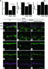
Figure 5
Figure 5. RS1 gene transfer restores photoreceptor calcium homeostasis and TRPM1 functionality in Rs1-KO retinas.
Rs1-KO retinas were injected with AAV8-scRS/IRBP-hRS1 viruses at P30 and examined at P92. At P92, compared with WT retinas, the untreated Rs1-KO retinas had a disrupted OPL with scattered signals of RIBEYE (purple, B and C) and low abundance of the TRPM1 channel (green, E and F) at the OPL, while treated Rs1-KO retinas revealed improved lamination of the OPL and the close proximity of the presynaptic ribbon to the TRPM1 channels at the ON-BC dendritic tips (D and G). (A) Functional rescue by RS1 gene transfer. Increases in rod photoreceptor synaptic free calcium concentration (untreated: 35% of that in WT; treated: 69% of that in WT [n = 4; *P < 0.05, Student’s t test]); TRPM1 intensity at ON-BC dendritic tips (percentage of whole-cell TRPM1 signal intensity; untreated: 37%; treated: 45% [n = 4; ***P < 0.001, Student’s t test]); and light-adapted RBC MP: untreated retina –37 ± 1.4 mV vs. treated retina –29.2 ± 1.2 mV (n = 4; ***P < 0.001, Student’s t test); WT retina –28.3 ± 1.4 mV. All Rs1-KO values were normalized to that of P30 WT. At P92, while VGluT1 (green) was localized at the OPL in WT retinas (H), it was scattered and less abundant at the disrupted OPL in untreated control retinas (I). Treatment led to a nearly normal appearance of VGluT1 immunoreactivity concentrated along the restored OPL of Rs1-KO retinas (J). Notably, the mGluR6 receptor (green) and RGS7 (purple) retained their normal localization at the ON-BC dendritic tips in Rs1-KO retinas regardless of the viral treatment (K and N: WT; L and O: untreated; M and P: AAV8-RS1 treated). Scale bars: 15 μm (BG); 20 μm (HP). n = 3.
Figure 4
Figure 4. RS1 expression in AAV8-scRS/IRBP-hRS1–injected and uninjected Rs1-KO mice at 2 months after treatment (P92).
Retinal sections from treated (right eye) and untreated (left eye) eyes of Rs1-KO mice 2 months after treatment, along with age-matched (P92) C57BL/6 WT mice were immunolabeled with anti-RS1 and anti-Gαo antibodies. RS1 gene transfer into Rs1-KO mice retina leads strong expression of RS1 protein (red) in inner segments (IS) of photoreceptors and OPL similar to that seen in WT mouse retina (A and C). Labeling was also prominent, but to a lesser extent, in ONL, INL, and IPL. In treated retinas, BCs staining with Gαo (green) revealed absence of schisis and much organized BC layer arrangement (F) as in WT retina (D), whereas the untreated retinas from Rs1-KO mice showed no RS1 labeling and displayed schisis cavities and BC disorganization (B and E). (GI) Gαo-RS1 overlay. Scale bars: 30 μm. n = 3.
Figure 3
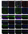
Figure 3. Deterioration of the mGluR6-signaling machinery in adolescent Rs1-KO mice.
Gαo, Gβ5, RGS7, and RGS11 are key components of the glutamate-activated mGluR6-signaling machinery, and they appeared to be normal in expression levels and protein localization at P22 (see Figure 2, A–H, and Supplemental Figure 1). At P30, compared with WT (A and E), VGluT1 (green) proteins are no longer concentrated at the Rs1-KO photoreceptor synaptic terminals (B and F). Meanwhile, in both WT and Rs1-KO retinas, RGS7 (red) proteins are concentrated at the dendritic tips of the BCs counterstained with PKCα antibody (blue), even though the OPL and BCs have been disorganized in the mutant retinas (WT: A, I, and M; Rs1-KO: B, J, and N). Costaining of RS1 (green), RGS11 (red), and Gβ5 (blue) showed that at P30, in WT retinas (C, G, K, and O), RGS11 and Gβ5 proteins are mostly distributed to the ON-BC dendritic tips, with minor portions at the cell somas, while RS1 proteins remain at dendritic shafts. (D, H, L, and P) RS1 proteins have been absent in Rs1-KO retinas. In addition to the normal dendritic tip localization, RGS11 and Gβ5 proteins are prominently expanded toward the soma and axon of the BCs in Rs1-KO retinas (L and P). Compared with WT (Q and R), Gαo proteins in Rs1-KO retinas (S and T) also showed a prominent localization at the ON-BC somas and axons. Scale bars: 20 μm. n = 3.
Figure 2
Figure 2. Photoreceptor-BC synaptic structure in WT and Rs1-KO retinas at P22.
(AH) En face image of whole-mounted WT (AD) and Rs1-KO (EH) retinas costained with antibodies against the presynaptic ribbon proteins RIBEYE (green) and Bassoon (red) and postsynaptic receptor protein mGluR6 (blue). Both WT and Rs1-KO retinas show normally intact rod photoreceptor active zones with horseshoe-shaped structures of RIBEYE and Bassoon, immediately adjacent to the mGluR6 receptor puncta specific for the dendritic tips of ON-BCs. At P22, Gαo proteins were normally concentrated at the dendritic tips and shafts (I, J), while Rs1-KO retinas had a grossly normal Gαo-staining pattern (K, L). Costaining of presynaptic VGluT1 (green), RGS7 (red) with PKCα (a marker for RBC; blue), and Gβ5 (green) with RGS11 (magenta) both showed similar patterns between WT (MP, U, V) and Rs1-KO (Q–T, W, X) retinas. Scale bars: 2 μm (AH); 20 μm (IX). n = 3.
Figure 1
Figure 1. Photoreceptor synaptic calcium deficiency and bipolar TRPM1 channel anomaly and hyperpolarized MP in P22 Rs1-KO retinas.
(AD) RS1 (green); counterstain Gαo (purple), a BC marker. In WT retina, Rs1 is profusely present in the photoreceptor inner segments and OPL. Rs1 is concentrated at the ON-BC dendritic shaft and sparsely present around the cell soma. (E and F) In Rs1-KO retinas at P22, presynaptic RIBEYE (blue) and CaV1.4 (red) are normally distributed as in WT. In contrast to WT retinas (G) where TRPM1 (green) proteins are aggregated at the dendritic tips opposing the presynaptic RIBEYE/CaV1.4 complex (I), the dendritic component of TRPM1 is greatly diminished (H, arrow, and J) in Rs1-KO retinas. (KL) At P22, mGluR6 (green) is exclusively located at the dendritic tips of the ON-BCs in both WT and Rs1-KO retinas. (M) At P22, measurements with Ca2+-sensitive dye fluo-4 showed that the median [Ca2+]i in Rs1-KO rod spherules was 48% of normalized median WT value (n = 7; ***P < 0.001; Student’s t test). (N) TRPM1 signal intensity at the bipolar dendritic tips was 36% of the whole-cell signal in Rs1-KO retinas, significantly lower than that of over 53% in WT (n = 5; ***P < 0.001; Student’s t test). (O) In line with the TRPM1 defects, the MP of light-exposed RBC is abnormally low in light-exposed Rs1-KO retinas (n = 5; ***P < 0.001; Student’s t test). GCL, ganglion cell layer. Scale bars: 10 μm (BD); 20 μm (A, EL). n = 4.

Comment in

References

    1. Jarraya B, et al. Dopamine gene therapy for Parkinson’s disease in a nonhuman primate without associated dyskinesia. Sci Transl Med. 2009;1(2):2ra4. - PubMed
    1. Alexander B, et al. Reversal of depressed behaviors in mice by p11 gene therapy in the nucleus accumbens. Sci Transl Med. 2010;2(54):54ra76. - PMC - PubMed
    1. Akil O, et al. Restoration of hearing in the VGLUT3 knockout mouse using virally mediated gene therapy. Neuron. 2012;75(2):283–293. doi: 10.1016/j.neuron.2012.05.019. - DOI - PMC - PubMed
    1. Sikkink SK, Biswas S, Parry NR, Stanga PE, Trump D. X-linked retinoschisis: an update. J Med Genet. 2007;44(4):225–232. doi: 10.1136/jmg.2006.047340. - DOI - PMC - PubMed
    1. Tantri A, et al. X-linked retinoschisis: a clinical and molecular genetic review. Surv Ophthalmol. 2004;49(2):214–230. doi: 10.1016/j.survophthal.2003.12.007. - DOI - PubMed

Publication types

MeSH terms