. 2015 Jul;22(7):522-31.
doi: 10.1038/nsmb.3051. Epub 2015 Jun 22.

Crystal structure, conformational fixation and entry-related interactions of mature ligand-free HIV-1 Env

Affiliations

Crystal structure, conformational fixation and entry-related interactions of mature ligand-free HIV-1 Env

Young Do Kwon et al. Nat Struct Mol Biol. 2015 Jul.

Abstract

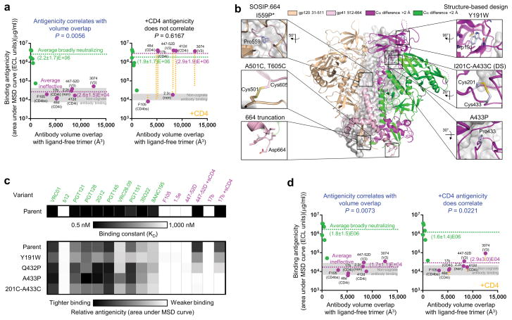
As the sole viral antigen on the HIV-1-virion surface, trimeric Env is a focus of vaccine efforts. Here we present the structure of the ligand-free HIV-1-Env trimer, fix its conformation and determine its receptor interactions. Epitope analyses revealed trimeric ligand-free Env to be structurally compatible with broadly neutralizing antibodies but not poorly neutralizing ones. We coupled these compatibility considerations with binding antigenicity to engineer conformationally fixed Envs, including a 201C 433C (DS) variant specifically recognized by broadly neutralizing antibodies. DS-Env retained nanomolar affinity for the CD4 receptor, with which it formed an asymmetric intermediate: a closed trimer bound by a single CD4 without the typical antigenic hallmarks of CD4 induction. Antigenicity-guided structural design can thus be used both to delineate mechanism and to fix conformation, with DS-Env trimers in virus-like-particle and soluble formats providing a new generation of vaccine antigens.


Conflict of interest statement

Competing financial interests The authors declare no competing financial interests.

Figures

Figure 1
Figure 1
Crystal structure of ligand-free HIV-1-Env trimer, and conformational changes related to individual subunit structures and virus entry. (a) The left gp120-gp41 protomer of the ligand-free BG505 SOSIP.664 trimer is shown in B-factor putty representation, the right protomer in Cα-backbone representation colored by average Cα distance between ligand-free Env and previously determined Env subunit structures, and the third protomer in grey cartoon representation. (b) Plot of pre-fusion Env B-factors versus Cα-subunit movement (see Supplementary Table 1 for a full listing of correlations and P-values for residue-level properties). (c) Correlations and P-values for pre-fusion B-factors of type 1 fusion machines versus pre-fusion to post-fusion movement of fusion subunit (see Supplementary Fig. 3 for subunit pictorials and correlation graphs). P-values in (b) and (c) were obtained by two-tailed student’s t test.
Figure 2
Figure 2
Ligand-free HIV-1-Env trimer is structurally compatible with epitopes of broadly neutralizing, but not ineffective, antibodies. (a) Superposition of ligand-free and antibody-bound HIV-1-Env structures. (Left) Ligand-free gp120 core monomer shown in ribbon representation, with regions of less (or greater) than 2 Å RMSD upon antibody binding shown in green (or magenta), and representative antibody-bound structures in gray. (Middle and right) Ligand-free and antibody-bound HIV-1-Env trimers. In the right panel, antibodies PGT122 and 35022 are shown in gray semitransparent surface, with rear protomer removed for clarity. RMSD values are reported in Supplementary Table 7. (b) Breadth-potency of broadly neutralizing (green) and ineffective (magenta) antibodies on a diverse 170 HIV-1-isolate panel. (c) Structural compatibility of ligand-free trimer by antibody epitope: an appropriate structural compatibility. The ligand-free Env structure is displayed as Cα-ribbon, with antibody epitope residues colored green (structurally compatible) or magenta (incompatible), and grey for non-epitope regions. RMSD (solid fill) and volume overlap (striped fill) with the respective antibody-Env complexes shown as a bar graph, with two linear scales split at RMSD and antibody-antigen volume overlap cutoffs of 2 and 500 Å3, respectively; bars below the respective cutoffs are colored green, and magenta otherwise. Antibody labels are colored green if the epitope is structurally compatible, magenta if incompatible, and gray if not present in the structure. (d) Ligand-free trimer structural compatibility versus antibody breadth. Volume overlap (left), RMSD (middle) and Antigenic Structural Compatibility (ASC) score (right) are graphed versus antibody breadth on a diverse 170 HIV-1-isolate panel; pictorial representations shown in Supplemental Fig. 5. P-values for Spearman correlations provided (n = 14).
Figure 3
Figure 3
Structural compatibility-guided negative selection and an appropriate target antigenicity. (a) Size-exclusion chromatography profile of crystallization-grade SOSIP before negative selection (left panel), and SDS-PAGE analyses of negative selection: first with antibody 447-52D, next with a cocktail of V3 antibodies, and third by CD4 (right panels). NR: non-reducing conditions, R: reducing conditions. (b) V3-epitope exposure on BG505 SOSIP.664 quantified by SPR on a panel of V3 antibodies and displayed as percent of total V3 epitope upon CD4 triggering. *V3 epitopes for antibodies 39F and 19b have not been structurally defined. (c) V3 antigenicity for all structurally defined antibodies in b mapped onto epitope atoms in the structure of the ligand-free Env trimer. (d) Appropriate target antigenicity. Binding antigenicity is displayed on the ligand-free Env trimer with epitope atoms with tight trimer binding to broadly neutralizing antibodies in green and to ineffective antibodies in magenta.
Figure 4
Figure 4
CD4-induced changes in antigenicity and conformational fixation of ligand-free HIV-1 Env. (a) BG505 SOSIP.664 ligand-free structural compatibility versus binding antigenicity, in the absence (left) and presence (right) of CD4 as measured by MSD. Antibodies are displayed in green (broadly neutralizing) and magenta (ineffective); the average binding of each is provided; and ineffective antibodies are labeled (CD4bs: CD4-binding site, CD4i: CD4-induced, non: non-neutralizing, V3: V3-loop directed). The change in binding to ineffective antibodies in the presence of CD4 is shown as a yellow dotted line. P-values for Spearman correlations are provided (n = 13). Binding levels for non-cognate antibodies lie within the gray shaded areas. (b) Conformational fixation of HIV-1-Env trimer. The central image depicts the ligand-free BG505 SOSIP.664 HIV-1-Env trimer, with two protomers shown in cartoon representation, one colored by domains (gp120 in wheat and gp41 in light pink), a second colored by RMSD distance between ligand-free trimer and subunit structures of CD4-bound gp120 and post-fusion gp41 (green if less than 2 Å, magenta if more than 2 Å). A third protomer is shown in gray. Insets: atomic-level details. *Residue 559 is disordered in the ligand-free structure. (c) Binding antigenicity of BG505 SOSIP.664 variants. Heat map showing binding of BG505 SOSIP.664 and variants that stabilized the ligand-free closed state to a panel of antibodies. (d) Ligand-free trimer structural compatibility versus BG505 SOSIP.664 201C-433C binding antigenicity in the absence (left) and presence (right) of CD4, with antibodies and average binding in green (broadly neutralizing) and magenta (ineffective) as in a. Elisa, Octet and Biacore data in Supplemental Data Set 2 and Supplementary Tables 5 and 6.
Figure 5
Figure 5
Atomic-level models and physical stability of ligand-free 201C-433C mutant (DS-SOSIP). (a) Atomic-level models of residues 201 and 433 in ligand-free pre-fusion closed state (grey) and CD4-bound state (yellow). Ribbon representations of the two structures are shown with residues that make-up the bridging sheet in the CD4-bound conformation colored orange, residues 201 and 433 in cyan spheres, and 201-433 Cα distance indicated. Variable loops are labeled. (Monomeric CD4-bound conformation modeled from PDB ID 2B4C, 3U4E and 3JWD). (b) Thermostability of the DS-SOSIP was assessed by differential scanning calorimetry. Raw data are shown in solid line (black for BG505 SOSIP.664 and red for DS-SOSIP), with corresponding curves from the fit shown in purple dashed lines. Tm values and error were obtained from the fit. (c) Physical stability of trimeric DS-SOSIP as determined by the quaternary-specific antibody VRC26.09 after 60 minutes of incubation at physical extremes or after 10 freeze-thaw cycles. Error bars are SEM of two technical replicates.
Figure 6
Figure 6
DS-SOSIP binds a single CD4 without the typical antigenic hallmarks of CD4 triggering. (a) Binding of soluble CD4 to SOSIP.664 and mutants measured by SPR with single-cycle kinetics. Values in parenthesis report standard errors from fit of the data to a 1:1 Langmuir binding model. Note that the level of binding for the P313W mutant is roughly 3-times higher than either wild-type SOSIP or 201C-433C. (b) Time course of CD4 activation of HIV-1 Env as measured by SPR. To initiate the time course, CD4 was mixed with HIV-1 Env at time point 0, and after incubation (time shown on x-axis), assessed by SPR for interaction with antibody (y-axis). (Left) Binding to antibody 17b, which recognizes a bridging sheet epitope; (right) binding to 3074, which recognizes a V3 epitope (right). (c) Sedimentation equilibrium analytical ultracentrifugation measurements of BG505 SOSIP.664 and 201C-433C variant in presence of excess 2-domain soluble CD4. Stochiometry for both 2-domain and 4-domain CD4 as well as residual calculation are provided in Supplementary Data Set 4.
Figure 7
Figure 7
An asymmetric intermediate in the HIV-1-entry pathway. (a) smFRET of JR-FL virions with and without 201C-433C substitution. Population FRET histograms are each paired with transition density plots, which display the relative density of observed transitions. Results for both ligand-free and dodecameric CD4 (sCD4D1D2-Igαtp), graphed in left and right pairs, respectively. The dominance of the low FRET state even in the presence of CD4 indicates DS-stabilized Env to remain in a closed state even when bound by CD4. (b) HIV-1 entry mechanism with conformation-blocking mutations, antigenicity, and interactions with functional ligands. A new mechanistic state, characterized by the binding of a single molecule of CD4 with no bridging sheet formation and reduced V3 exposure, is highlighted in red. Env density from refs. and .
Figure 8
Figure 8
A new generation of conformationally fixed HIV-1-Env trimeric immunogens. (a) HDX characterization of conformational mobility. Changes in exchange of hydrogen-deuterium relative to wild-type BG505 SOSIP.664 are displayed, with regions that become more ordered (blue) or disordered (red) shown on one lobe of the ligand-free trimer. Full HDX profiles are shown in Supplementary Fig. 7. (b) Antigenic characteristics of SOS virus-like particles (VLPs, comprising SOS mutant without IP) from strain JR-FL (modified with E168K to allow binding of V1V2-directed broadly neutralizing antibodies) and strain BG505 (modified with T332N to allow binding of 2G12 antibody). While broadly neutralizing antibody binding is maintained between parent and 201C-433C VLPs, the 201C-433C variant showed reduced ineffective antibody binding, especially in the presence of CD4. Broadly neutralizing antibodies binding are shown in green, ineffective in magenta and binding of antibodies in presence of 2 μg/ml of sCD4 in orange. Ineffective antibodies labeled (CD4bs: CD4-binding site, CD4i: CD4-induced, V3: V3-loop directed). (c) Information flow of antigenicity-guided immunogen design: from human antibody response through structure and antigenicity-guided design to conformationally fixed immunogens.

References

    1. Starcich BR, et al. Identification and characterization of conserved and variable regions in the envelope gene of HTLV-III/LAV, the retrovirus of AIDS. Cell. 1986;45:637–48. - PubMed
    1. Wei X, et al. Antibody neutralization and escape by HIV-1. Nature. 2003;422:307–12. - PubMed
    1. Chen L, et al. Structural basis of immune evasion at the site of CD4 attachment on HIV-1 gp120. Science. 2009;326:1123–7. - PMC - PubMed
    1. Kwong PD, et al. HIV-1 evades antibody-mediated neutralization through conformational masking of receptor-binding sites. Nature. 2002;420:678–82. - PubMed
    1. Fouts TR, Binley JM, Trkola A, Robinson JE, Moore JP. Neutralization of the human immunodeficiency virus type 1 primary isolate JR-FL by human monoclonal antibodies correlates with antibody binding to the oligomeric form of the envelope glycoprotein complex. J Virol. 1997;71:2779–85. - PMC - PubMed

Publication types

Associated data