Review
. 2015:44:311-37.
doi: 10.1146/annurev-biophys-051013-023008.

Structural Symmetry in Membrane Proteins

Affiliations
Review

Structural Symmetry in Membrane Proteins

Lucy R Forrest. Annu Rev Biophys. 2015.

Abstract

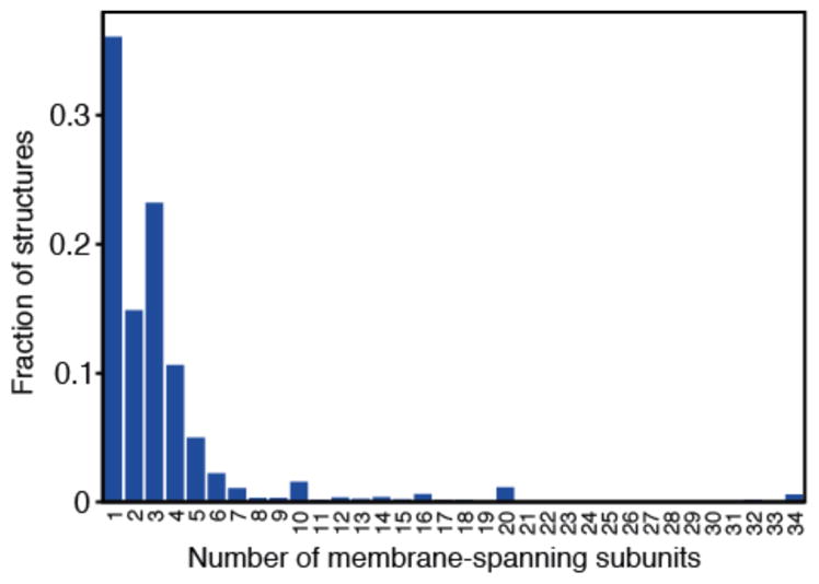
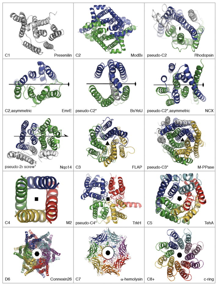
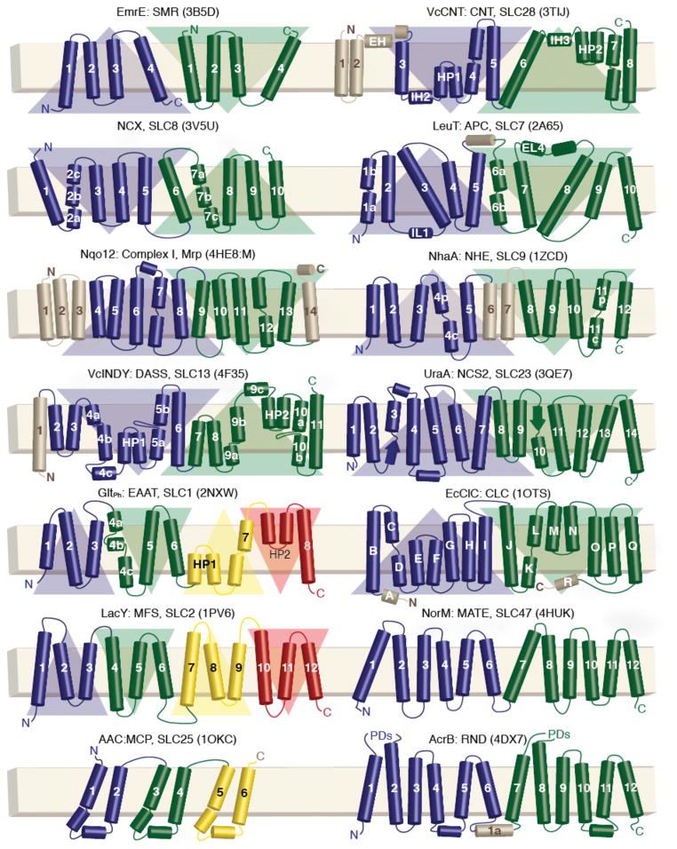
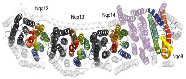
Symmetry is a common feature among natural systems, including protein structures. A strong propensity toward symmetric architectures has long been recognized for water-soluble proteins, and this propensity has been rationalized from an evolutionary standpoint. Proteins residing in cellular membranes, however, have traditionally been less amenable to structural studies, and thus the prevalence and significance of symmetry in this important class of molecules is not as well understood. In the past two decades, researchers have made great strides in this area, and these advances have provided exciting insights into the range of architectures adopted by membrane proteins. These structural studies have revealed a similarly strong bias toward symmetric arrangements, which were often unexpected and which occurred despite the restrictions imposed by the membrane environment on the possible symmetry groups. Moreover, membrane proteins disproportionately contain internal structural repeats resulting from duplication and fusion of smaller segments. This article discusses the types and origins of symmetry in membrane proteins and the implications of symmetry for protein function.

Keywords: alternating access; asymmetry; internal repeats; inverted-topology repeats; oligomer.


Figures

Figure 1
Figure 1
Degree of oligomer formation in membrane protein structures. Data was taken from the PDB_TM database (103) dated 2014.08.01 containing 2241 proteins. No filtering of low-resolution models or redundancy was applied. Thus, each data-point may contain a number of representatives from the same structural family. This analysis is necessarily biased toward those proteins that crystallize and are well studied. Oligomers are formed via interactions between membrane domains as well as between fused water-soluble domains.
Figure 2
Figure 2
Point symmetry types in membrane protein structures. Structures are shown as cartoon helices, viewed down onto the membrane. Different colors are used to indicate symmetric elements, i.e., independent chains or (*)internal repeats. Non-symmetric elements are in gray. EmrE is an asymmetric homo-dimer (PDB entry: 3B5D), while other transporters are asymmetric as well as pseudo-symmetric (e.g., NCX, 3V5U). Two-fold screw axis (22)-pseudo-symmetry is seen in the Mrp antiporter-like subunits of complex I (see Figure 4). Presenilin is an aspartate protease (4HYG). ModB2 is from the homodimeric molybdate type I ABC importer (2ONK). MalFG is from the heterodimeric MalFGK2 type I ABC importer (2R6G). BsYetJ is a pH-dependent Ca2+ channel from the TM Bax inhibitor motif (TMBIM) family (4PGW). FLAP, or five-lipooxygenase-activating protein, is a member of the family of membrane-associated proteins in eicosanoid and glutathione metabolism (MAPEG, 2Q7M). M-PPase is a membrane pyrophosphatase (4AV3). M2 is a H+ channel from influenza A (3LBW, TM domains only). TrkH is a K+ channel from the superfamily of K+ transporters (SKT, 3PJZ). TehA is a SLAC anion channel homolog (3M73 chain A). Connexin-26 is a gap junction (2ZW3); each hemi-channel exhibits C6 symmetry. α-hemolysin is a pore-forming toxin (7AHL). The c8-ring is a F-type ATP synthase membrane rotor (2XND). Symmetry axes were defined using SymD v1.3 (52) and figures were made with Pymol v1.7 (Schrödinger Ltd).
Figure 3
Figure 3
Transmembrane topologies of secondary transporters of known structure. The outside of the cell or organelle is oriented to the top. Protein name, family name, human solute carrier (SLC) nomenclature, and representative PDB identifiers (in parentheses) are given. Helices are represented as cylinders, and strands as arrows. Each inverted-topology repeat is highlighted using a triangle whose base is on the side of the N-terminus. Abbreviations: Vc, Vibrio cholerae; CNT, concentrative nucleoside transporter; APC, amino acid/polyamine/organocation superfamily; AAC, ADP-ATP carrier; PDs, periplasmic domains.
Figure 4
Figure 4
The membrane domain of complex I from T. thermophilus (PDB entry 4HE8). Inverted-topology repeats related by a two-fold (22) screw-axis are colored blue to red for the first repeat or dark gray for the second repeat. The single ‘repeat’ in subunit Nqo8 has same TM topology as the three ‘first repeats’, but is rotated by 180° around an axis perpendicular to the membrane plane (cf. dark blue helices), and is more tilted. Nqo8 is separated from Nqo14 by a number of additional subunits (purple). Other non-symmetric segments and subunits are colored white.

References

    1. Abraham A-L, Pothier J, Rocha EPC. Alternative to Homo-oligomerisation: The Creation of Local Symmetry in Proteins by Internal Amplification. J Mol Biol. 2009;394(3):522–34. - PubMed
    1. Almén M, Nordström KJ, Fredriksson R, Schiöth HB. Mapping the human membrane proteome: a majority of the human membrane proteins can be classified according to function and evolutionary origin. BMC Biol. 2009;7(1):50. - PMC - PubMed
    1. André I, Strauss CEM, Kaplan DB, Bradley P, Baker D. Emergence of symmetry in homooligomeric biological assemblies. Proc Natl Acad Sci USA. 2008;105(42):16148–52. - PMC - PubMed
    1. Arabidopsis Genome Initiative. Analysis of the genome sequence of the flowering plant Arabidopsis thaliana. Nature. 2000;408(6814):796–815. - PubMed
    1. Arthur CP, Stowell MHB. Structure of Synaptophysin: A Hexameric MARVEL-Domain Channel Protein. Structure. 2007;15(6):707–14. - PMC - PubMed

Publication types

Substances

LinkOut - more resources