. 2015 Jul;17(7):893-906.
doi: 10.1038/ncb3192. Epub 2015 Jun 22.

Molecular characterization of LC3-associated phagocytosis reveals distinct roles for Rubicon, NOX2 and autophagy proteins

Affiliations

Molecular characterization of LC3-associated phagocytosis reveals distinct roles for Rubicon, NOX2 and autophagy proteins

Jennifer Martinez et al. Nat Cell Biol. 2015 Jul.

Retraction in

Abstract

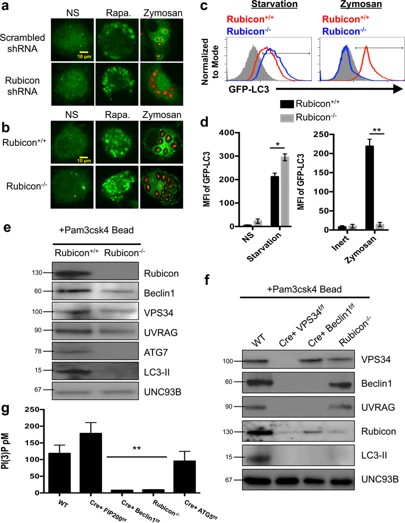
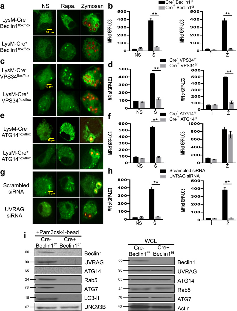
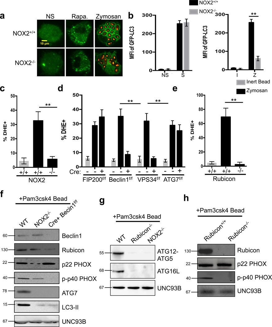
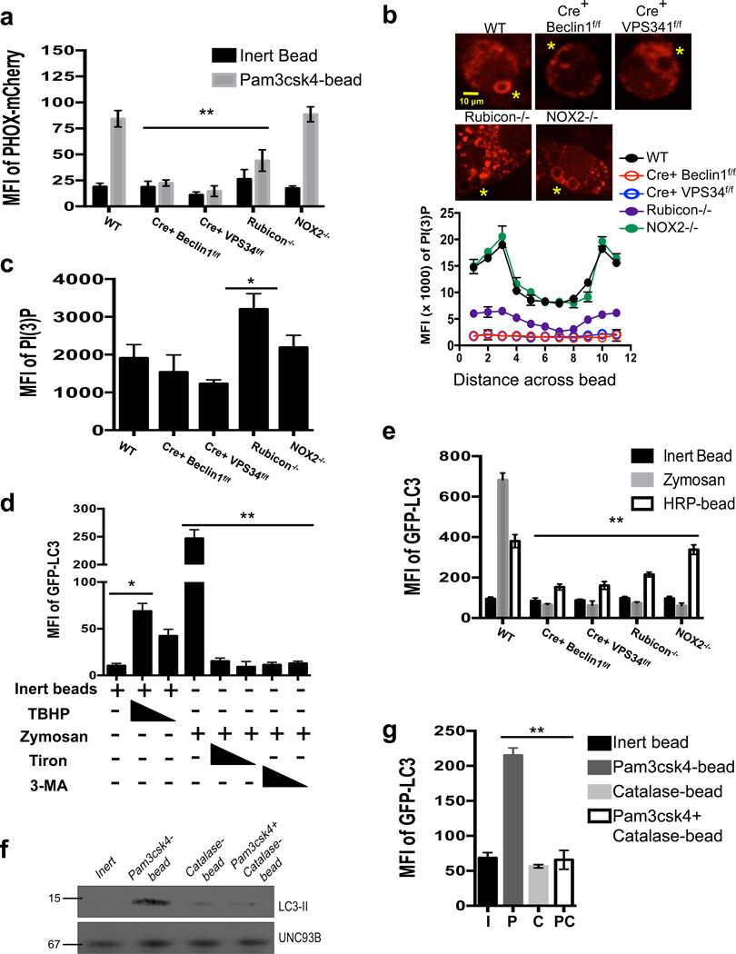
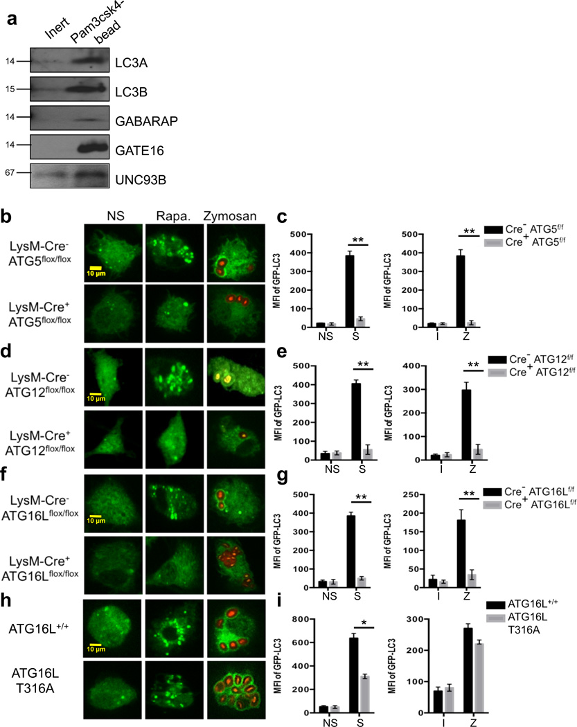
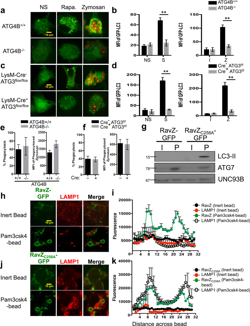
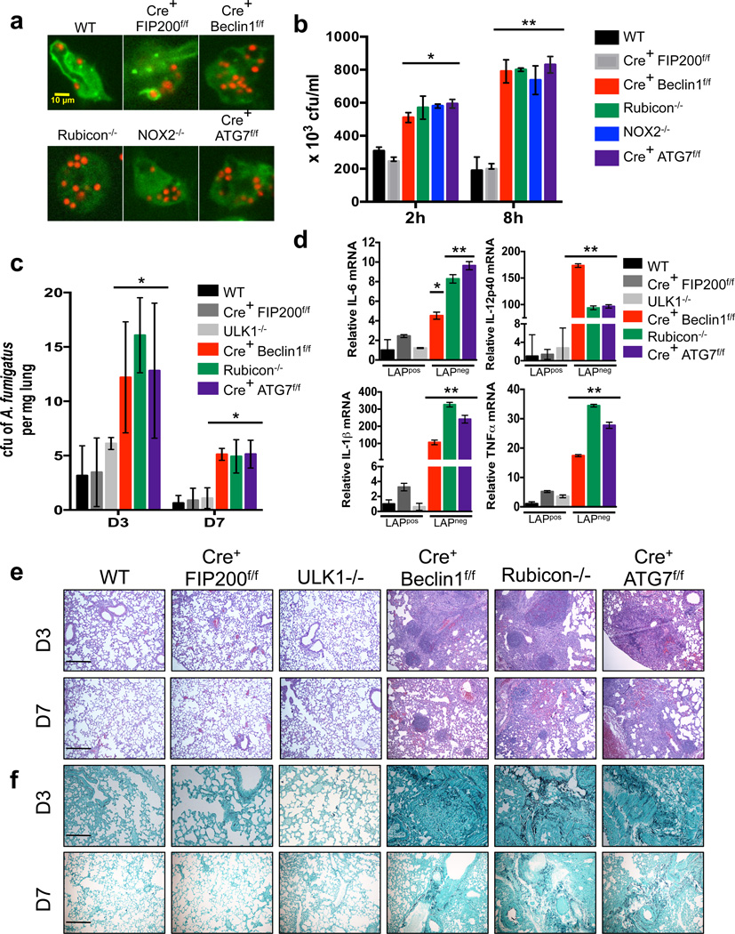
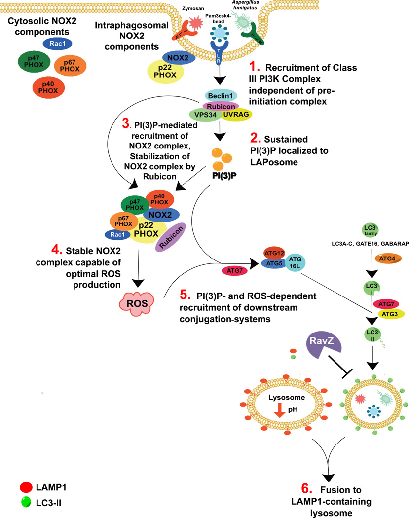
LC3-associated phagocytosis (LAP) is a process wherein elements of autophagy conjugate LC3 to phagosomal membranes. We characterize the molecular requirements for LAP, and identify Rubicon as being required for LAP but not autophagy. Rubicon is recruited to LAPosomes and is required for the activity of a Class III PI(3)K complex containing UVRAG but lacking ATG14 and Ambra1. This allows for the sustained localization of PtdIns(3)P, which is critical for recruitment of downstream autophagic proteins and stabilization of the NOX2 complex to produce reactive oxygen species. Both PtdIns(3)P and reactive oxygen species are required for conjugation of LC3 to LAPosomes and subsequent association with LAMP1(+) lysosomes. LAP is induced by engulfment of Aspergillus fumigatus, a fungal pathogen that commonly afflicts immunocompromised hosts, and is required for its optimal clearance in vivo. Therefore, we have identified molecules that distinguish LAP from canonical autophagy, thereby elucidating the importance of LAP in response to A. fumigatus infection.


Figures

Figure 1
Figure 1. Rubicon is required for LAP
(a) RAW-GFP-LC3 cells were transduced with lentiviral constructs for Scrambled shRNA or Rubicon shRNA. At 72 hours after transfection, cells were left untreated (NS) or treated with 200 nM rapamycin (Rapa.) or fed Alexa Fluor 594-zymosan (Zymosan). GFP-LC3 puncta was assessed at 18 h, and translocation of GFP-LC3 to the LAPosome was assessed at 1 h. (b–d) Rubicon+/+ GFP-LC3+ and Rubicon−/− GFP-LC3+ bone marrow-derived macrophages were left untreated (NS) or were cultured with 200 nM rapamycin (Rapa., b), starvation conditions (c), or Alexa Fluor 594-zymosan (Zymosan, b–c). GFP-LC3 puncta was assessed at 18 h, and translocation of GFP-LC3 to the LAPosome was assessed at 1 h by confocal microscopy (b) and flow cytometry (c). Filled grey histogram represents inert bead. (d) The mean fluorescence intensity (MFI) of membrane bound GFP-LC3 was determined using flow cytometry under starvation conditions (left) and zymosan feeding (right). Data are presented as mean ± SD of three independent experiments (*p < 0.05, **p < 0.001). (e–f) Bone marrow-derived macrophages from Rubicon+/+ and Rubicon−/− mice (e) or WT, LysM-Cre+ VPS34flox/flox, LysM-Cre+ Beclin1flox/flox, and Rubicon−/− mice (f) were allowed to phagocytose latex beads coated with Pam3csk4 for 1 hr. Phagosomes were purified using sucrose gradient as described in experimental procedures. Phagosome proteins were solubilized in SDS-PAGE and blotted with the indicated antibodies. The results presented are representative of three independent experiments. (g) Bone marrow-derived macrophages from genetic knockout strains were fed Pam3csk4-beads (30 minutes). mVPS34 was immunoprecipitated from the purified LAPosomes, and equivalent amounts were used in the Class III PI3K Activity assay. Data, as pM of PI(3)P, is presented as mean ± SD of three independent experiments (**p < 0.001).
Figure 2
Figure 2. LAP utilizes a UVRAG-containing Class III PI3K Complex
(a–b) Bone marrow-derived macrophages from LysM-Cre− Beclin1flox/flox GFP-LC3+ and LysM-Cre+ Beclin1flox/flox GFP-LC3+ mice were left untreated (NS) or were cultured with 200 nM rapamycin (Rapa., a), starvation conditions (S, b), Inert beads (I, b), or Alexa Fluor 594-zymosan (Zymosan or Z, a–b). (c–d) Bone marrow-derived macrophages from LysM-Cre− VPS34flox/flox GFP-LC3+ and LysM-Cre+ VPS34flox/flox GFP-LC3+ mice were left untreated (NS) or were cultured with 200 nM rapamycin (Rapa., c), starvation conditions (S, d), Inert beads (I, d), or Alexa Fluor 594-zymosan (Zymosan or Z, c–d). (e–f) Bone marrow-derived macrophages from LysM-Cre− ATG14flox/flox and LysM-Cre+ ATG14flox/flox mice were transfected with GFP-LC3. After 48 hours of transfection, cells were left untreated (NS) or were cultured with 200 nM rapamycin (Rapa., e), starvation conditions (S, f), Inert beads (I, f), or Alexa Fluor 594-zymosan (Zymosan or Z, e–f). (g–h) RAW-GFP-LC3 cells were transfected with Scrambled or UVRAG siRNA oligonucleotides. After 48 hours of transfection, cells were left untreated (NS) or were cultured with 200 nM rapamycin (Rapa., g), starvation conditions (S, h), Inert beads (I, h), or Alexa Fluor 594-zymosan (Zymosan or Z, g–h). GFP-LC3 puncta was assessed at 18 h, and translocation of GFP-LC3 to the LAPosome was assessed at 1 h by confocal microscopy (a, c, e, g) and flow cytometry (b, d, f, h). Data are presented as mean ± SD of three independent experiments (**p < 0.001). (i) Bone marrow-derived macrophages from LysM-Cre− Beclin1flox/flox and LysM-Cre+ Beclin1flox/flox mice were allowed to phagocytose latex beads coated with Pam3csk4 for 1 hr. Phagosomes were purified using sucrose gradient as described in experimental procedures. Phagosome proteins (left), as well as whole cell lysates from non-stimulated cells (right), were solubilized in SDS-PAGE and blotted with the indicated antibodies. The results presented are representative of three independent experiments.
Figure 3
Figure 3. NOX2 is downstream of the Class III PI3K Complex and required for LAP
(a–b) Bone marrow-derived macrophages from NOX2+/+ GFP-LC3+ and NOX2−/− GFP-LC3+ mice were left untreated (NS) or were cultured with 200 nM rapamycin (Rapa., a), starvation conditions (S, b), Inert beads (I, b), or Alexa Fluor 594-zymosan (Zymosan or Z, a–b). GFP-LC3 puncta was assessed at 18 h, and translocation of GFPLC3 to the LAPosome was assessed at 1 h by confocal microscopy (a) and flow cytometry (b). (c–e) Bone marrow-derived macrophages from genetic knockout strains were fed inert beads or Alexa Fluor 594-zymosan and analyzed for ROS production at 1 h by flow cytometry using dihydroethidium (DHE). Data are presented as mean ± SD of three independent experiments (**p < 0.001). (f–h) Bone marrow-derived macrophages from WT, NOX2−/−, and LysM-Cre+ Beclin1flox/flox mice (f), Rubicon+/+ and Rubicon−/− mice (g), and WT, Rubicon−/−, and NOX2−/− mice (h) were allowed to phagocytose latex beads coated with Pam3csk4 for 1 hr. Phagosomes were purified using sucrose gradient as described in experimental procedures. Phagosome proteins were solubilized in SDS-PAGE and blotted with the indicated antibodies. The results presented are representative of three independent experiments.
Figure 4
Figure 4. PI(3)P and ROS are both required for LAP
(a–c) Bone marrow-derived macrophages from genetic knockout strains were transiently transfected with PX-mCherry. After 48 hours of transfection, cells were fed inert beads or Pam3csk4-beads for 30 minutes. PI(3)P on LAPosomes was analyzed by flow cytometry (a) or confocal microscopy (b–c). Yellow asterisks indicate internalized beads. Representative images and signal intensity profiles for PX-mCherry across phagocytosed beads are quantified and shown graphically (n ≥ 25 / genotype) (b). Masks were generated around whole cells and the MFI of PI(3)P within the cell was quantified using Slidebook software. Data are presented as mean ± SD of three independent experiments (**p < 0.001) (c). Data are presented as mean ± SD of two independent experiments (n ≥ 25 / genotype). (d) RAW-GFP-LC3 cells were fed inert beads or zymosan, in the presence or absence of tert-butyl hydroperoxide (TBHP, 100 µM, 50 µM), Tiron (1 mM, 0.5 mM), or 3-MA (25 mM, 5 mM). Translocation of GFP-LC3 to the LAPosome was assessed at 1 h by flow cytometry. (e) Bone marrow-derived macrophages from GFP-LC3+ genetic knockout strains were fed inert beads, Alexa Fluor 594-zymosan, or HRP-coupled beads. Translocation of GFP-LC3 to the LAPosome was assessed at 1 h by flow cytometry. Data are presented as mean ± SD of three independent experiments (**p < 0.001) (f–g) RAW-GFP-LC3 cells were fed inert beads (I), Pam3csk4-beads (P), Catalase-beads (C), or Pam3csk4+Catalase-beads (PC). Translocation of GFP-LC3 to the LAPosome was assessed at 1 h by immunoblot analysis of purified phagosomal proteins (f) and flow cytometry (g). Data are presented as mean ± SD of three independent experiments (**p < 0.001).
Figure 5
Figure 5. The ATG5-12-16L and LC3-PE conjugation systems are required for LAP
(a) RAW cells were allowed to phagocytose inert beads or Pam3csk4-beads for 1 hour. Phagosomes were purified using sucrose gradient as described in experimental procedures. Phagosome proteins (left), as well as whole cell lysates from non-stimulated cells (right), were solubilized in SDS-PAGE and blotted with the indicated antibodies. The results presented are representative of three independent experiments. (b–c) Bone marrow-derived macrophages from LysM-Cre− ATG5flox/flox GFP-LC3+ and LysM-Cre+ ATG5flox/flox GFP-LC3+ mice were left untreated (NS) or were cultured with 200 nM rapamycin (Rapa., b), starvation conditions (S, c), Inert beads (I, c), or Alexa Fluor 594-zymosan (Zymosan or Z, b–c). (d–e) Bone marrow-derived macrophages from LysM-Cre− ATG12flox/flox and LysM-Cre+ ATG12flox/flox mice were transfected with GFP-LC3. After 48 hours of transfection, cells were left untreated (NS) or were cultured with 200 nM rapamycin (Rapa., d), starvation conditions (S, e), Inert beads (I, e), or Alexa Fluor 594-zymosan (Zymosan or Z, d–e). (f–g) Bone marrow-derived macrophages from LysM-Cre− ATG16Lflox/flox GFP-LC3+ and LysM-Cre+ ATG16Lflox/flox GFP-LC3+ mice were left untreated (NS) or were cultured with 200 nM rapamycin (Rapa., f), starvation conditions (S, g), Inert beads (I, g), or Alexa Fluor 594-zymosan (Zymosan or Z, f–g). (h–i) Bone marrow-derived macrophages from ATG16L+/+ and ATG16L T316A mice were transfected with GFP-LC3. After 48 hours of transfection, cells were left untreated (NS) or were cultured with 200 nM rapamycin (Rapa., h), starvation conditions (S, i), Inert beads (I, i), or Alexa Fluor 594-zymosan (Zymosan or Z, h–i). GFP-LC3 puncta was assessed at 18 h, and translocation of GFP-LC3 to the LAPosome was assessed at 1 h by confocal microscopy (b, d, f, h) and flow cytometry (c, e, g, i). (j) ATG4B+/+ and ATG4B−/− macrophages were fed Alexa Fluor 594-zymosan, and the percent of phagocytosis (% Phagocytosis, left) and the extent of phagocytosis (MFI of Phagocytosed Zymosan, right) was quantified by flow cytometry. (k) LysM-Cre− ATG3flox/flox and LysM-Cre+ ATG3flox/flox macrophages were fed Alexa Fluor 594-zymosan, and the percent of phagocytosis (% Phagocytosis, left) and the extent of phagocytosis (MFI of Phagocytosed Zymosan, right) was quantified by flow cytometry. Data are presented as mean ± SD of three independent experiments (**p < 0.001).
Figure 6
Figure 6. LAP-induced phagosome maturation requires LC3-II
(a–b) Bone marrow-derived macrophages from ATG4B+/+ and ATG4B−/− mice were transfected with GFP-LC3. After 48 hours of transfection, cells were left untreated (NS) or were cultured with 200 nM rapamycin (Rapa., a), starvation conditions (S, b), Inert beads (I, b), or Alexa Fluor 594-zymosan (Zymosan or Z, a–b). (c–d) Bone marrow-derived macrophages from LysM-Cre− ATG3flox/flox and LysM-Cre+ ATG3flox/flox mice were transfected with GFP-LC3. After 48 hours of transfection, cells were left untreated (NS) or were cultured with 200 nM rapamycin (Rapa., c), starvation conditions (S, d), Inert beads (I, d), or Alexa Fluor 594-zymosan (Zymosan or Z, c–d). GFP-LC3 puncta was assessed at 18 h, and translocation of GFP-LC3 to the LAPosome was assessed at 1 h by confocal microscopy (a, c) and flow cytometry (b, d). Data are presented as mean ± SD of three independent experiments (**p < 0.001). (e–h) RAW cells were transfected with RavZ-GFP (e–f) or RavZC258A-GFP (g–h). After 48 hours of transfection, cells were fed inert beads or Pam3csk4-beads for 1 hour. Immunofluorescent staining was performed for LAMP1 and analyzed by microscopy. Representative images (e, g) and signal intensity profiles (f, h) for RavZ, RavZC258A, and LAMP1 across phagocytosed beads are quantified and shown graphically (n ≥ 20 / genotype). Data are presented as mean ± SD of two independent experiments. (i) RAW cells were transfected with RavZ-GFP or RavZC258A-GFP. After 48 hours of transfection, cells were allowed to phagocytose inert beads or Pam3csk4-beads for 1 hour. Phagosomes were purified using sucrose gradient as described in experimental procedures. Phagosome proteins (left), as well as whole cell lysates from non-stimulated cells (right), were solubilized in SDS-PAGE and blotted with the indicated antibodies. The results presented are representative of three independent experiments.
Figure 7
Figure 7. Clearance of Aspergillus fumigatus requires LAP
(a) Bone marrow-derived macrophages from GFP-LC3+ genetic knockout strains were fed live A. fumigatus-dsRed at an MOI of 5, and translocation of GFP-LC3 to the LAPosome was assessed at 1 h by confocal microscopy. Representative images are shown (n ≥ 20 / genotype). (b) Bone marrow-derived macrophages from different genetic knockout strains were fed live A. fumigatus at an MOI of 1, and colony-forming units per ml (cfu/ml) of cell lysate was measured at 2 and 8 hours post-infection. Data are presented as mean ± SD of three independent experiments (*p< 0.05, **p < 0.01). (c–f) Mice of different genetic knockout strains were infected intranasally with live A. fumigatus conidia. A. fumigatus cfu per mg of lung tissue was measured on days 3 and 7 post-infection (c). Relative mRNA levels of IL-6, IL-1β, IL-12p40, and TNFα in infected lung tissue homogenates at day 3 post-infection were quantified by real-time PCR. Data normalized to actin. Data are presented as mean ± SD of three independent experiments (*p< 0.05). Lung histopathology [H&E staining, (e) and Gomorri staining (f)] on days 3 and 7 post-infection. Representative images are shown of 10× magnification. Scale bars represent 100 µm.
Figure 8
Figure 8. Proposed Model of LC3-Associated Phagocytosis
Recruitment of the Rubicon- and UVRAG-containing Class III PI3K complex allows for sustained VPS34 activity at the LAPosome, resulting in significant PI(3)P deposition on the LAPosome membrane. This PI(3)P allows for the recruitment of autophagic downstream conjugation systems to the LAPosome and stabilizes the NOX2 complex via its binding to p-p40PHOX. Rubicon itself also stabilizes the NOX2 complex, promoting optimal ROS production. Both PI(3)P and ROS are required for the recruitment of the downstream conjugation systems. LAP requires the activity of ATG5-ATG12-ATG16L complex, as well as ATG3 and ATG4, all of which are critical to the lipidation of LC3. Importantly, the maturation of the LAPosome requires the presence of LC3-II, as macrophages that express the Legionella effector protease, RavZ, fail to mature into LAMP1+ LAPosomes.

Comment in

References

    1. Levine B. Eating oneself and uninvited guests: autophagy-related pathways in cellular defense. Cell. 2005;120:159–162. - PubMed
    1. de Luca A, et al. IL-1 receptor blockade restores autophagy and reduces inflammation in chronic granulomatous disease in mice and in humans. Proc Natl Acad Sci U S A. 2014;111:3526–3531. - PMC - PubMed
    1. Levine B, Mizushima N, Virgin HW. Autophagy in immunity and inflammation. Nature. 2011;469:323–335. - PMC - PubMed
    1. Klionsky DJ, et al. Guidelines for the use and interpretation of assays for monitoring autophagy. Autophagy. 2012;8:445–544. - PMC - PubMed
    1. Sanjuan MA, et al. Toll-like receptor signalling in macrophages links the autophagy pathway to phagocytosis. Nature. 2007;450:1253–1257. - PubMed

Publication types

MeSH terms