. 2015 Jun 22;10(6):e0130249.
doi: 10.1371/journal.pone.0130249. eCollection 2015.

Combining α-Radioimmunotherapy and Adoptive T Cell Therapy to Potentiate Tumor Destruction

Affiliations

Combining α-Radioimmunotherapy and Adoptive T Cell Therapy to Potentiate Tumor Destruction

Jérémie Ménager et al. PLoS One. .

Abstract

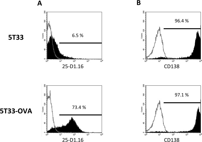
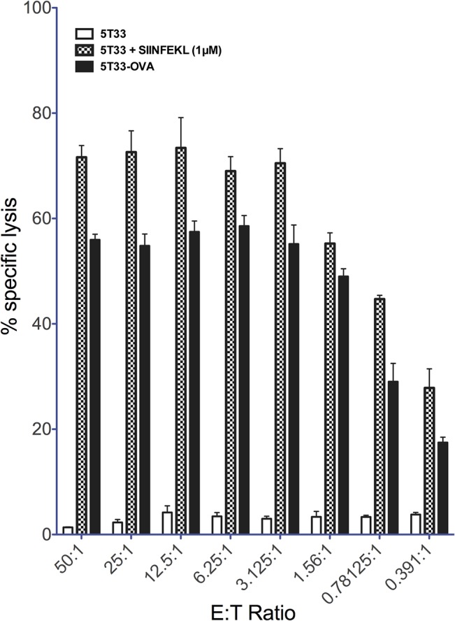
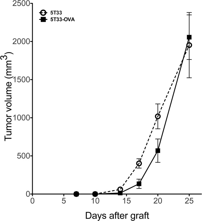
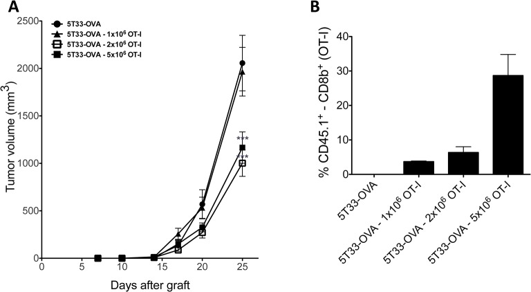
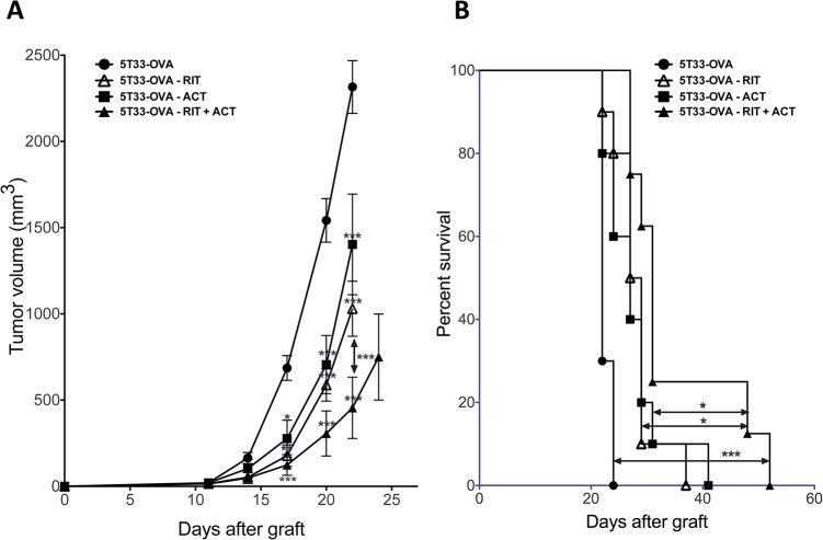
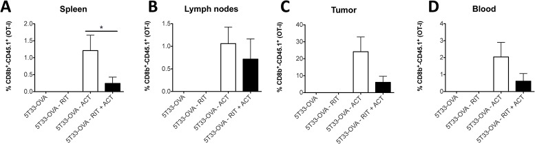
Ionizing radiation induces direct and indirect killing of cancer cells and for long has been considered as immunosuppressive. However, this concept has evolved over the past few years with the demonstration that irradiation can increase tumor immunogenicity and can actually favor the implementation of an immune response against tumor cells. Adoptive T-cell transfer (ACT) is also used to treat cancer and several studies have shown that the efficacy of this immunotherapy was enhanced when combined with radiation therapy. α-Radioimmunotherapy (α-RIT) is a type of internal radiotherapy which is currently under development to treat disseminated tumors. α-particles are indeed highly efficient to destroy small cluster of cancer cells with minimal impact on surrounding healthy tissues. We thus hypothesized that, in the setting of α-RIT, an immunotherapy like ACT, could benefit from the immune context induced by irradiation. Hence, we decided to further investigate the possibilities to promote an efficient and long-lasting anti-tumor response by combining α-RIT and ACT. To perform such study we set up a multiple myeloma murine model which express the tumor antigen CD138 and ovalbumine (OVA). Then we evaluated the therapeutic efficacy in the mice treated with α-RIT, using an anti-CD138 antibody coupled to bismuth-213, followed by an adoptive transfer of OVA-specific CD8+ T cells (OT-I CD8+ T cells). We observed a significant tumor growth control and an improved survival in the animals treated with the combined treatment. These results demonstrate the efficacy of combining α-RIT and ACT in the MM model we established.


Conflict of interest statement

Competing Interests: The authors declare that the research was conducted in the absence of any commercial or financial relationships that could be construed as a potential conflict of interest.

Figures

Fig 1
Fig 1. 5T33-OVA phenotypic analysis after transduction by a lentiviral vector encoding cytoplasmic ovalbumin (A) 5T33 and 5T33-OVA staining with PE-conjugated antibody 25-D1.16, which specifically recognizes the OVA peptide ‘SIINFEKL’ bound to the MHC class I molecule H-2Kb and (B) staining of 5T33 and 5T33-OVA with APC-conjugated anti-mouse CD138 mAb.
Flow cytometry was performed on a BD FACSCalibur Flow Cytometry System.
Fig 2
Fig 2. In vitro recognition of 5T33-OVA.
Cytotoxic T-cell-mediated lysis of 5T33-OVA targets. T cells isolated from OT-I mice were activated using irradiated splenocytes loading with OVA257–264 (SIINFEKL) peptide. In vitro T-cell-mediated cytotoxicity against 5T33-OVA was determined using a standard four-hour 51Cr release assay at several effector-to-target ratios (E:T Ratio).
Fig 3
Fig 3. In vivo tumor growth of 5T33-OVA.
Growth comparison of established tumors 5T33 versus 5T33-OVA. Animals were injected subcutaneously with 2x10⁶ tumor cells (n = 5 mice per group).
Fig 4
Fig 4. Adoptive OT-I T cell transfer.
(A), Dose response of 5T33-OVA tumor cells to adoptive transfer of OT-I CD8+ T cells treatment. Animals were injected subcutaneously with 2x10⁶ tumor cells and received the indicated doses of OT-I CD8+ T cells (n = 10 mice per group). Tumor volume was determined by using a caliper. Data points represent mean ± SD of 10 measures. *** p<0,001 as determined by two-way ANOVA and Bonferonni post-tests. (B), Tumors were explanted and single cell suspensions were prepared by grinding tumors in a tissu grinder. Cells were stained with monoclonal anti-CD8b PE and anti-CD45.1 FITC. Histograms represent the percentage of CD45.1+ in CD8+ cells.
Fig 5
Fig 5. Combination of RIT with adoptive immunotherapy inhibits tumor growth and prolongs survival.
(A), Animals were injected with 2x10⁶ tumor cells subcutaneously and received the indicated treatment (n = 8 to 10 mice per group). Tumor volume was measured. Data points represent mean ± SD. * p<0,05, ** p<0,01, *** p<0,001 as determined by two-way ANOVA and Bonferonni post-tests. The combination of RIT + ACT or RIT alone or the ACT transfer alone significantly decrease tumor growth when compared with the control. The combination of RIT + ACT significantly decrease tumor growth when compared RIT alone or the ACT transfer alone. (B), Animals were injected subcutaneously with 2x10⁶ tumor cells and received the indicated treatment. The percentage of surviving mice was evaluated when tumor volume reach end-point of 2500 mm3. The combination of RIT + ACT significantly increased survival (median survival of 31 days; log-rank, 0.0001) when compared with control or RIT alone (median survival of 28 days; log-rank, 0,0413) or the ACT transfer alone (median survival of 27 days; log-rank, 0,0391) cohorts. Statistical analysis were performed using non-parametric Mann-Whitney test.
Fig 6
Fig 6. OT-I CD8+ T cells persist within the tumor but also in the lymphoid organs and the periphery.
After the different treatment as indicated on the graph (n = 8 to 10 per group), when tumour reached a volume of 2500 mm3, the mice were sacrificed and spleen (A), lymph nodes (B), tumor (C) and blood (D) were harvested. Then, cells were stained with monoclonal anti-CD8b PE and anti-CD45.1 FITC. Histograms represent the percentage of CD45.1+ in CD8+ cells. Statistical analysis were performed using non-parametric Mann-Whitney test.

References

    1. Delaney G, Jacob S, Featherstone C, Barton M (2005) The role of radiotherapy in cancer treatment: estimating optimal utilization from a review of evidence-based clinical guidelines. Cancer 104: 1129–1137. 10.1002/cncr.21324 - DOI - PubMed
    1. Begg AC, Stewart FA, Vens C (2011) Strategies to improve radiotherapy with targeted drugs. Nat Rev Cancer 11: 239–253. 10.1038/nrc3007 - DOI - PubMed
    1. Bernier J, Hall EJ, Giaccia A (2004) Timeline: Radiation oncology: a century of achievements. Nat Rev Cancer 4: 737–747. 10.1038/nrc1451 - DOI - PubMed
    1. Mothersill C, Seymour CB (2004) Radiation-induced bystander effects—implications for cancer. Nat Rev Cancer 4: 158–164. 10.1038/nrc1277 - DOI - PubMed
    1. Prise KM, O'Sullivan JM (2009) Radiation-induced bystander signalling in cancer therapy. Nat Rev Cancer 9: 351–360. 10.1038/nrc2603 - DOI - PMC - PubMed

Publication types