. 2015 Aug;18(8):1109-15.
doi: 10.1038/nn.4049. Epub 2015 Jun 22.

Subtype-specific plasticity of inhibitory circuits in motor cortex during motor learning

Affiliations

Subtype-specific plasticity of inhibitory circuits in motor cortex during motor learning

Simon X Chen et al. Nat Neurosci. 2015 Aug.

Abstract

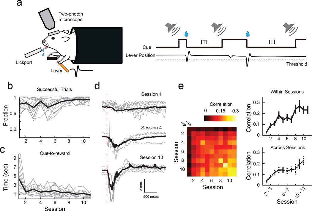
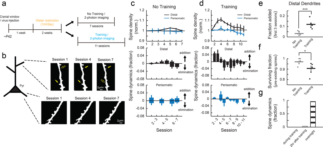
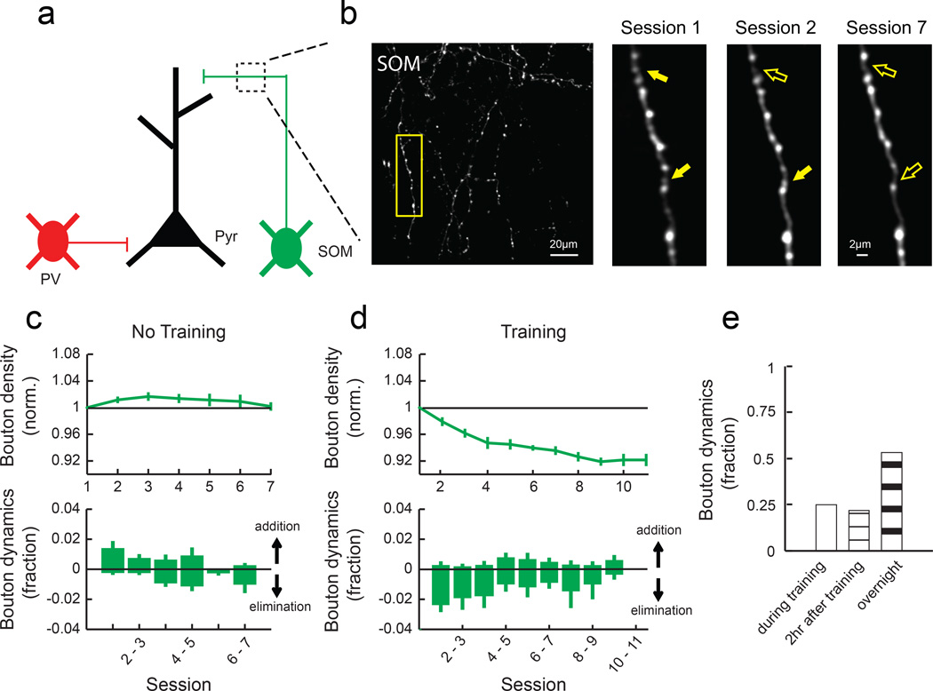
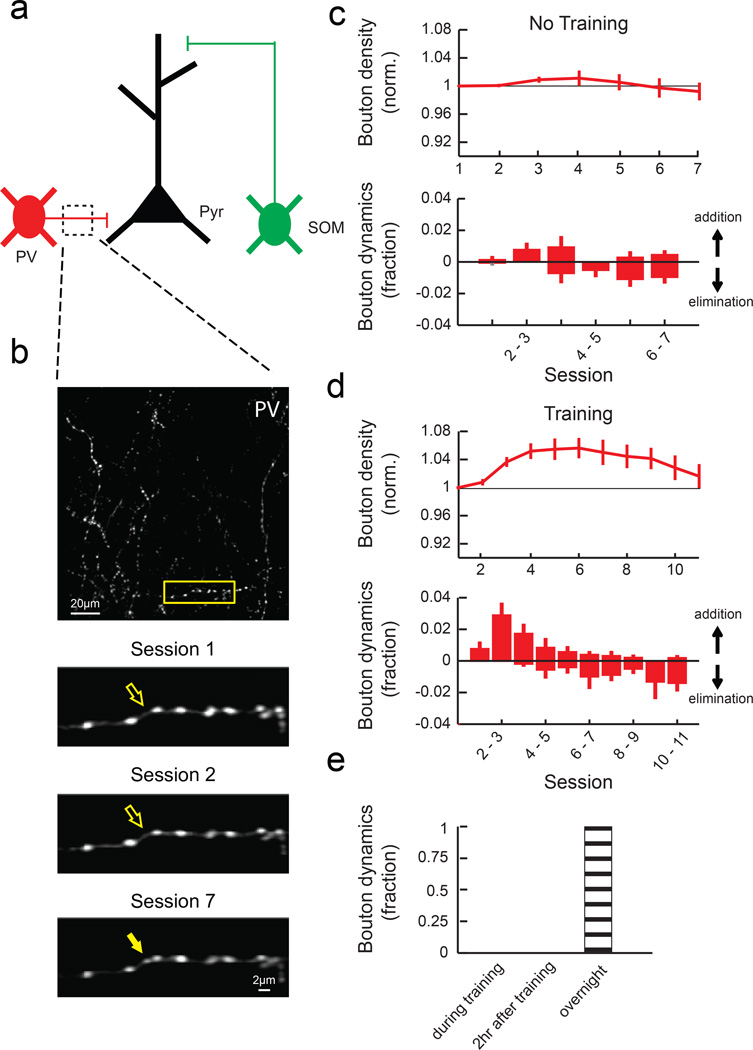
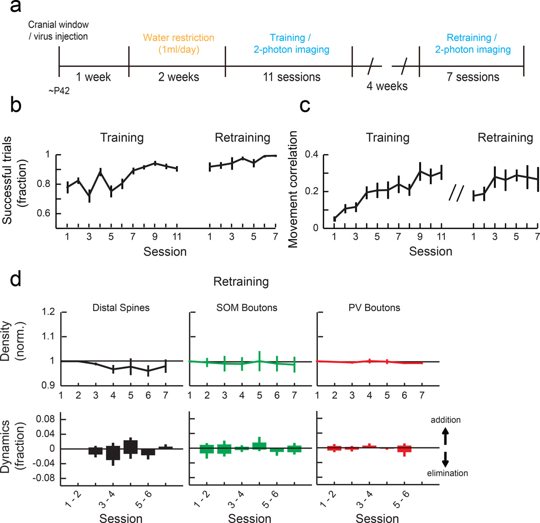
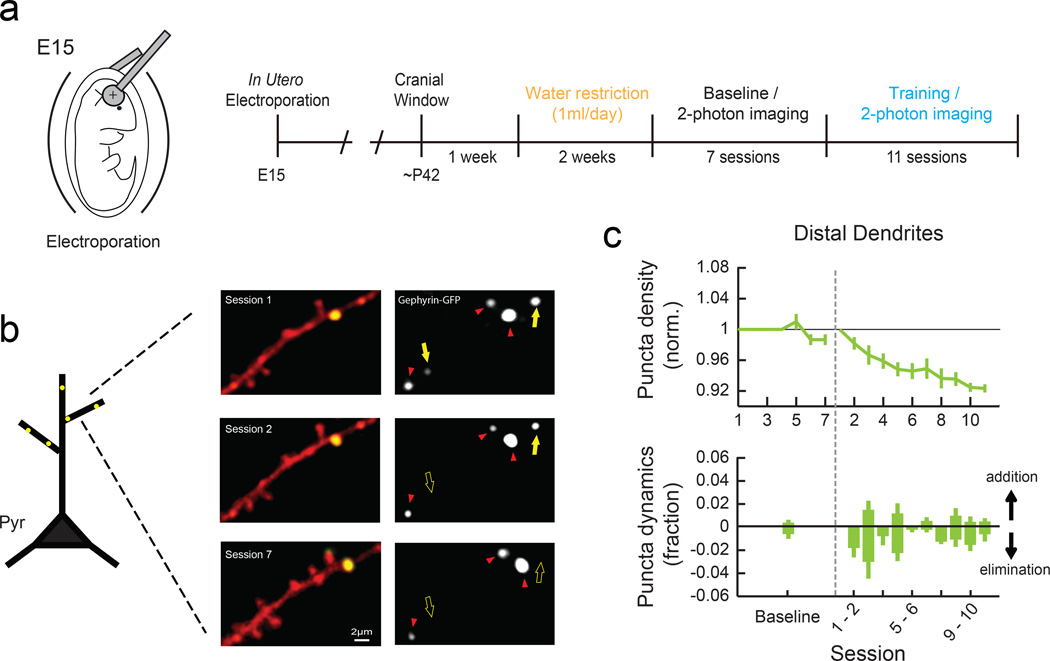
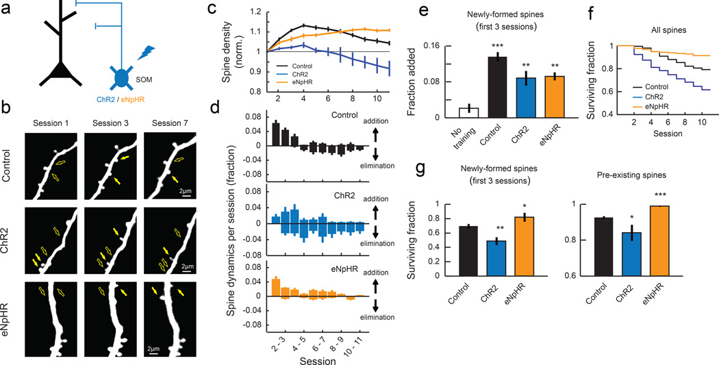
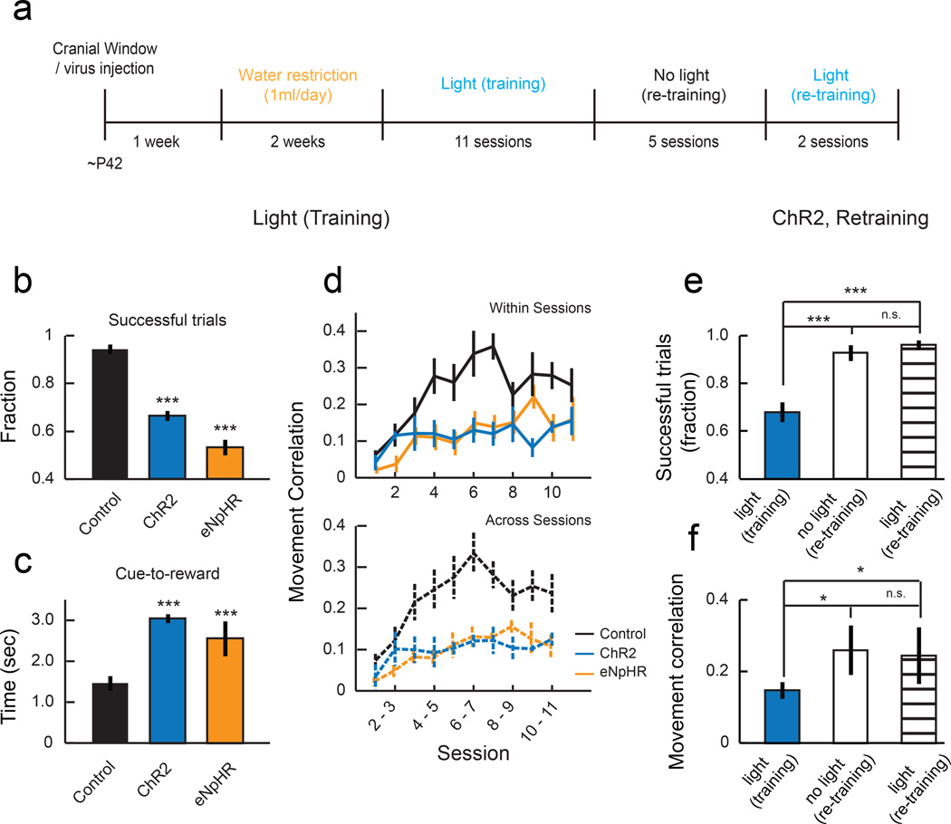
Motor skill learning induces long-lasting reorganization of dendritic spines, principal sites of excitatory synapses, in the motor cortex. However, mechanisms that regulate these excitatory synaptic changes remain poorly understood. Here, using in vivo two-photon imaging in awake mice, we found that learning-induced spine reorganization of layer (L) 2/3 excitatory neurons occurs in the distal branches of their apical dendrites in L1 but not in the perisomatic dendrites. This compartment-specific spine reorganization coincided with subtype-specific plasticity of local inhibitory circuits. Somatostatin-expressing inhibitory neurons (SOM-INs), which mainly inhibit distal dendrites of excitatory neurons, showed a decrease in axonal boutons immediately after the training began, whereas parvalbumin-expressing inhibitory neurons (PV-INs), which mainly inhibit perisomatic regions of excitatory neurons, exhibited a gradual increase in axonal boutons during training. Optogenetic enhancement and suppression of SOM-IN activity during training destabilized and hyperstabilized spines, respectively, and both manipulations impaired the learning of stereotyped movements. Our results identify SOM inhibition of distal dendrites as a key regulator of learning-related changes in excitatory synapses and the acquisition of motor skills.


Figures

Fig. 1
Fig. 1. Lever-press task for head-fixed mice
(a) Schematic of experimental setup and task. (b) Fraction of successful trials improves with learning (P<0.001, 1-way ANOVA, n = 17 mice). Grey, individual animals; black, mean. (c) Time from cue onset to reward decreases with learning (P<0.001, 1-way ANOVA). Grey, medians of individual animals; black, mean. (d) Example lever movement traces from one animal aligned by movement onset, showing the emergence of movement stereotypy with learning. Grey, 10 individual trials; black, median of all trials; red dotted line, movement onset. (e), Left, trial-to-trial correlation of movement kinematics during learning. Each square represents the median value of the pairwise correlations of the rewarded movement traces of all trial pairs within the session pair, averaged across animals. Right, movement correlation (movement stereotypy) increases within and across sessions, corresponding to the diagonals shown by the solid and dotted arrows in the correlogram on the left (within sessions, P<0.001; across sessions, P<0.001, 1-way ANOVA, n = 17 mice).
Fig. 2
Fig. 2. Motor learning induces compartment-specific reorganization of dendritic spines
(a) Experimental timeline. (b) Repeated imaging of the distal portion of apical dendrites (L1, 0 – 50 μm from pia) and perisomatic dendrites (L2/3, 150 – 200 μm) of L2/3 pyramidal cells throughout learning. Filled and open arrows indicate present and absent dynamic spines, respectively. (c) Mean spine density normalized to the initial session (top) and daily spine dynamics (bottom) of distal dendrites (n = 7 mice, 269 spines) and perisomatic dendrites (n = 4 mice, 120 spines) in no training animals. (d) Mean spine density normalized to the initial session (top) and daily spine dynamics (bottom) of distal dendrites (n = 5 mice, 251 spines) and perisomatic dendrites (n = 5 mice, 206 spines) in training animals. Learning transiently increases spine density in distal but not perisomatic dendrites of L2/3 pyramidal cells (Distal: P<0.001, compared to no training; Perisomatic: P=0.246, compared to no training, 2-way ANOVA). (e) Learning increases the spine addition rate in the distal dendrites during the first 3 sessions. (f) Learning increases the elimination rate of pre-existing spines in the distal dendrites (fraction of pre-existing spines that remained until session 7). *P<0.05, ***P<0.001, one-tailed bootstrap test. (g) Spine formation and elimination in the distal dendrites rarely occurred during (0%, 0/25) or within two hours (4%, 1/25) of training sessions (n = 3 mice, 134 spines total). Error bars indicate SEM.
Fig. 3
Fig. 3. SOM-IN axonal boutons are rapidly eliminated following training
(a) SOM-INs mainly inhibit distal dendrites of excitatory neurons. (b) Left, a field of SOM-IN axons in L1 imaged throughout learning in vivo. Right, zoom of outlined area on the left. Filled and open arrows indicate present and absent dynamic boutons, respectively. (c) Mean normalized bouton density (top) and daily bouton dynamics (bottom) of SOM-INs in no training animals (n = 6 mice, 464 boutons). (d) Mean normalized bouton density (top) and daily bouton dynamics (bottom) of SOM-INs in training animals (n = 5 mice, 433 boutons). SOM boutons decreased with training (P<0.001, 2-way ANOVA with post hoc Tukey's test, compared to ʽno trainingʼ) due to an increase in bouton elimination (P<0.001, one-tailed bootstrap). (e) Many bouton formation and elimination events of SOM-INs occurred during (25%, 8/32) and within two hours (22%, 7/32) of training sessions (n = 3 mice, 258 boutons).
Fig. 4
Fig. 4. PV-IN axonal boutons transiently increase during learning
(a) PV-INs mainly inhibit perisomatic regions. (b) Top, a field of PV-IN axons in L2/3. Bottom, zoom of outlined area above. (c) Mean normalized bouton density (top) and daily bouton dynamics (bottom) of PV-INs in no training animals (n = 6 mice, 488 boutons). (d) Mean normalized bouton density (top) and daily bouton dynamics (bottom) of PV-INs in training animals (n = 5 mice, 396 boutons). PV boutons increase with training (P<0.001, 2-way ANOVA with post hoc Tukey's test, compared to ʽno trainingʼ). (e) Bouton formation and elimination of PV-INs did not occur during or within two hours of training sessions (n = 3 mice, 12 dynamic boutons out of 215 total boutons). Error bars indicate SEM.
Fig. 5
Fig. 5. Synaptic reorganization is not observed during performance of a previously learned task
(a) Experimental timeline for retraining animals. (b) Fraction of successful trials in training (n = 17 mice) and retraining mice (n = 10 mice). (c) Median pairwise correlation of rewarded movements within each session in training and retraining mice. (d) Mean normalized density of distal spines, SOM boutons and PV boutons (top) and their daily dynamics in each session (bottom) during retraining (Spine: n = 3 mice, 181 spines, SOM: n = 3 mice, 196 boutons, PV: n = 3 mice, 254 boutons). The density of spines and boutons showed no significant changes (Spine: P=0.63, SOM boutons: P=0.99, PV boutons: P=0.91, 1-way ANOVA). Error bars indicate SEM.
Fig. 6
Fig. 6. Elimination of inhibitory synapses in the distal dendrites of L2/3 pyramidal neurons during learning
(a) Schematic of in utero electroporation to express Gephyrin-GFP in neocortical L2/3 pyramidal neurons (left) and experimental timeline (right). (b) Left, Representative images of a distal dendritic branch in red with Gephyrin-GFP puncta shown in green. Right, the Gephyrin-GFP channel only. Yellow filled and open arrows indicate present and absent dynamic puncta, respectively. Red arrowheads indicate stable puncta. (c) Mean normalized Gephyrin-GFP puncta density (top) and daily dynamics (bottom) during baseline (7 sessions, n = 3 mice, 138 puncta) and learning (11 sessions, n = 4 mice, 339 puncta). For baseline, puncta dynamics from all sessions are combined. Black dotted line represents the beginning of the behavioral training. Gephyrin-GFP puncta are reduced with training (P<0.001, 2-way ANOVA with post hoc Tukey's test, compared to baseline) due to an increase in puncta elimination compared to the baseline (P<0.001, one-tailed bootstrap). Error bars indicate SEM.
Fig. 7
Fig. 7. Manipulation of SOM-IN activity during training disrupts spine stability
(a) ChR2 or eNpHR was expressed to activate or inactivate SOM-INs during training sessions, respectively. tdTomato was expressed in control animals. (b) Repeated imaging of L1 distal dendritic branches of excitatory neurons of control, ChR2, and eNpHR animals throughout learning. Filled and open arrows indicate present and absent dynamic spines, respectively. (c) Mean normalized spine density in control animals (ʽControlʼ, n = 12 mice, 665 spines), animals in which SOM-INs were activated during training (ʽChR2ʼ, n = 5 mice, 255 spines), and animals in which SOM-INs were inactivated during training (ʽeNpHRʼ, n = 6 mice, 397 spines). SOM-IN activation blocks learning-related increase of spine density (P<0.001, 2-way ANOVA with post hoc Tukey's test, compared to control), and SOM-IN inactivation extends the spine density increase (P<0.001, 2-way ANOVA with post hoc Tukey's test, compared to control). (d) Daily spine dynamics in control, ChR2, and eNpHR animals during training. (e) Training-induced spine formation in the first 3 sessions of control, ChR2, and eNpHR animals. **P<0.01, ***P<0.001, one-tailed bootstrap with Bonferroni correction compared to ʽNo trainingʼ. (f) Kaplan-Meier survival curves of all dendritic spines. Spines are less stable when SOM-INs are activated and more stable when SOM-INs are inactivated compared to control (P<0.001, log-rank test with Bonferroni correction). (g) Left, fraction of newly-formed spines in the first 3 sessions of training that remained until the end of the training. SOM-IN activation reduced the stability of learning-related new spines whereas SOM-IN inactivation hyperstabilized them. Right, fraction of pre-existing spines that remained until the end of training. *P<0.05, **P<0.01, ***P<0.001, one-tailed bootstrap test with Bonferroni correction compared to ʽControlʼ.
Fig. 8
Fig. 8. Manipulation of SOM-IN activity impaired the formation of stereotyped movements
(a) Experimental timeline. (b) Mean fractions of successful trials in sessions 7–11, showing that control animals achieved a reward in a larger fraction of trials than ChR2 or eNpHR animals. ***P<0.001, one-tailed bootstrap test with Bonferroni correction compared to ʽControlʼ. (c) Time from cue onset to achieve reward is longer in ChR2 and eNpHR animals compared to control. ***P<0.001, one-tailed bootstrap test with Bonferroni correction compared to ʽControlʼ. (d) Medians of trial-trial movement correlations. Values are lower in ChR2 and eNpHR animals, indicating their failure to form stereotyped movement patterns (within sessions: P<0.001; across sessions: P<0.001, compared to ʽControlʼ, 2-way ANOVA with post hoc Tukey's test). Error bars indicate SEM. (e) Mean fractions of successful trials of ChR2 animals in the last 2 sessions of training with light, retraining without light, and retraining with light (n = 5 mice). (f) Mean correlation of movements within sessions in all 3 conditions. Once the animals acquire the motor skill, SOM-IN stimulation did not impact the performance (P>0.1, ‘no light (re-training)’ vs. ‘light (retraining)’). *P<0.05, ***P<0.001, one-tailed bootstrap test with Bonferroni correction). Error bars indicate SEM.

Comment in

  • Removing synaptic brakes on learning.
    Grillo FW, West L, De Paola V. Grillo FW, et al. Nat Neurosci. 2015 Aug;18(8):1062-4. doi: 10.1038/nn.4073. Nat Neurosci. 2015. PMID: 26216460 No abstract available.

References

    1. Peters AJ, Chen SX, Komiyama T. Emergence of reproducible spatiotemporal activity during motor learning. Nature. 2014;510:263–267. - PubMed
    1. Costa RM, Cohen D, Nicolelis MA. Differential corticostriatal plasticity during fast and slow motor skill learning in mice. Curr Biol. 2004;14:1124–1134. - PubMed
    1. Huber D, et al. Multiple dynamic representations in the motor cortex during sensorimotor learning. Nature. 2012;484:473–478. - PMC - PubMed
    1. Nudo RJ, Milliken GW, Jenkins WM, Merzenich MM. Use-dependent alterations of movement representations in primary motor cortex of adult squirrel monkeys. J Neurosci. 1996;16:785–807. - PMC - PubMed
    1. Rioult-Pedotti MS, Friedman D, Donoghue JP. Learning-induced LTP in neocortex. Science. 2000;290:533–536. - PubMed

Publication types