Comparative Study
. 2015 Jun 23:10:23.
doi: 10.1186/s13024-015-0017-8.

Nigral overexpression of alpha-synuclein in the absence of parkin enhances alpha-synuclein phosphorylation but does not modulate dopaminergic neurodegeneration

Affiliations
Comparative Study

Nigral overexpression of alpha-synuclein in the absence of parkin enhances alpha-synuclein phosphorylation but does not modulate dopaminergic neurodegeneration

Anne-Sophie Van Rompuy et al. Mol Neurodegener. .

Abstract

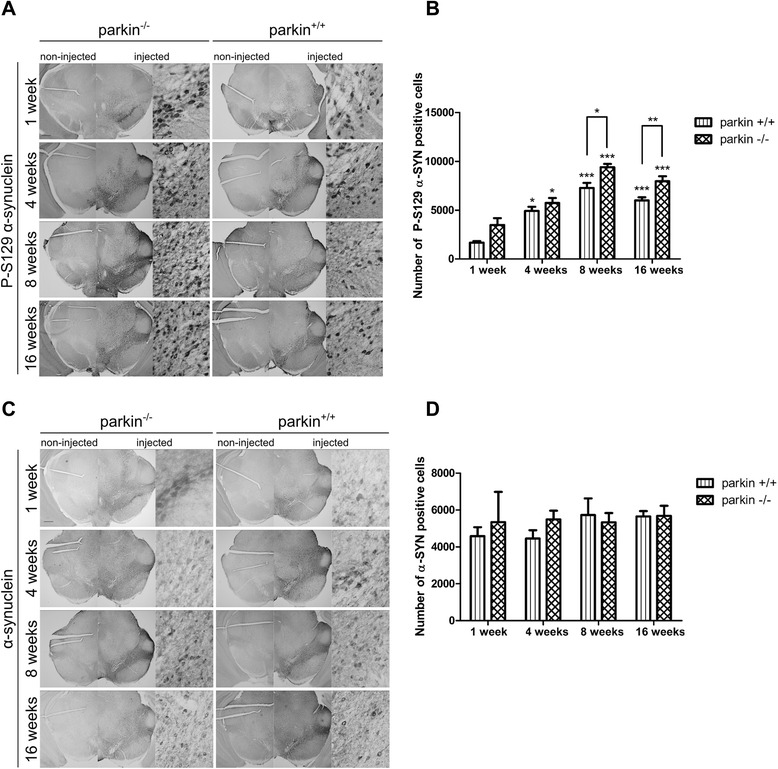
Background: Alpha-synuclein is a key protein in the pathogenesis of Parkinson's disease. Mutations in the parkin gene are the most common cause of early-onset autosomal recessive Parkinson's disease, probably through a loss-of-function mechanism. However, the molecular mechanism by which loss of parkin function leads to the development of the disease and the role of alpha-synuclein in parkin-associated Parkinson's disease is still not elucidated. Conflicting results were reported about the effect of the absence of parkin on alpha-synuclein-mediated neurotoxicity using a transgenic approach. In this study, we investigated the effect of loss of parkin on alpha-synuclein neuropathology and toxicity in adult rodent brain using viral vectors. Therefore, we overexpressed human wild type alpha-synuclein in the substantia nigra of parkin knockout and wild type mice using two different doses of recombinant adeno-associated viral vectors.

Results: No difference was observed in nigral dopaminergic cell loss between the parkin knockout mice and wild type mice up to 16 weeks after viral vector injection. However, the level of alpha-synuclein phosphorylated at serine residue 129 in the substantia nigra was significantly increased in the parkin knockout mice compared to the wild type mice while the total expression level of alpha-synuclein was similar in both groups. The increased alpha-synuclein phosphorylation was confirmed in a parkin knockdown cell line.

Conclusions: These findings support a functional relationship between parkin and alpha-synuclein phosphorylation in rodent brain.


Figures

Figure 1
Figure 1
Overexpression of α-SYN or eGFP in mouse SN with high titer rAAV2/7 vectors. Immunohistochemical staining for TH (left panels) shows cell loss in the injected side of both the parkin−/− and the parkin+/+ mice at 4 weeks after injection with a high titer of rAAV2/7-WT α-SYN. Immunohistochemical stainings for α-SYN and eGFP (middle and right panels) show wide expression of these proteins in the injected side of the SN. Right panels are magnifications of the adjacent left panel. Scale bars overviews = 400 μm and magnifications = 50 μm.
Figure 2
Figure 2
High transduction efficiency of mouse dopaminergic neurons with rAAV2/7-WT α-SYN and rAAV2/7-eGFP. Fluorescent double stainings for TH and α-SYN at 1 week or TH and eGFP at 4 weeks after injection of rAAV2/7-WT α-SYN or rAAV2/7-eGFP respectively, demonstrate that the majority of the dopaminergic neurons of the injected side of the SN is transduced. Scale bar = 25 μm.
Figure 3
Figure 3
A high dose of rAAV2/7-WT α-SYN induces similar dopaminergic degeneration in parkin−/− and parkin+/+ mice. Stereological quantification of the number of TH-positive neurons in the SN of parkin+/+ and parkin−/− mice at (A) 1 week and 4 weeks after injection with a high titer of rAAV2/7-WT α-SYN and at (B) 4 weeks after injection with rAAV2/7-eGFP. (C) quantification of TH staining in striatum (injected over non-injected side) of parkin+/+ and parkin−/− mice 4 weeks after injection. Asterisks depict significant decrease respective to 1 week, unless specified otherwise. (Mean ± SEM, two-way ANOVA followed by Bonferroni post-hoc test, ** p < 0.01 ***p < 0.001, 1 week rAAV2/7-WT α-SYN: n = 4; 4 weeks rAAV2/7-WT α-SYN: n = 15-16; 4 weeks rAAV2/7-eGFP: n = 5-6).
Figure 4
Figure 4
Increased phosphorylation of α-SYN at S129 in parkin−/− mice compared to parkin+/+ mice. (A) Representative images of P-S129 α-SYN expression in the SN of parkin−/− and parkin+/+ mice at 1 week and 4 weeks after injection with a high titer of rAAV2/7-WT α-SYN. Right panels are magnifications of the overviews of the injected side (middle panels). Scale bar overviews = 400 μm and magnifications = 50 μm. (B) Stereological quantification of the number of P-S129 α-SYN positive cells in the injected side of the SN of parkin+/+ and parkin−/− mice at 1 week (n = 4) and 4 weeks (n = 15-16) after injection with a high titer of rAAV2/7-WT α-SYN. (Mean ± SEM, two-way ANOVA followed by Bonferroni post-hoc test, # p < 0.05 versus parkin+/+, **p < 0.01 versus 1 week, ***p < 0.001 versus 1 week). (C) Stereological quantification of the number of α-SYN positive cells in the injected side of the SN of parkin+/+ and parkin−/− mice at 1 week (n = 4) and 4 weeks (n = 15-16) after injection with a high titer of rAAV2/7-WT α-SYN. (Mean ± SEM). (D) Quantification of the number of P-S129 α-SYN positive neuritic inclusions in the striatum at 4 weeks (n = 14–15, Mean ± SEM). (E) Picture of the P-S129 α-SYN staining in the striatum (left: 10 x) and magnification (right: 40 x) of neuritic inclusions. Scale bar = 50 μm.
Figure 5
Figure 5
A low dose of rAAV2/7-WT α-SYN induces similar dopaminergic degeneration in parkin−/− and parkin+/+ mice. (A) Representative images of immunohistochemical staining for TH in the SN at 1 week, 4 weeks, 8 weeks and 16 weeks after injection with a low titer of rAAV2/7-WT α-SYN. Scale bar = 400 μm. (B) Stereological quantification of the number of TH-positive neurons in the SN of parkin+/+ and parkin−/− mice at 1 week, 4 weeks, 8 weeks and 16 weeks after injection with a low titer of rAAV2/7-WT α-SYN. (Mean ± SEM, two-way ANOVA followed by Bonferroni post-hoc test, *p < 0.05, **p < 0.01, n = 2-3 at 1 week, n = 10-13 at 4 weeks, n = 5-6 at 8 weeks, n = 6 at 16 weeks).
Figure 6
Figure 6
Increased P-S129 α-SYN in parkin−/− mice after injection with a low titer of rAAV2/7-WT α-SYN. (A) Representative images of P-S129 α-SYN expression in the SN of parkin−/− and parkin+/+ mice at 1 week, 4 weeks, 8 weeks and 16 weeks after injection with a low titer of rAAV2/7-WT α-SYN. Right panels are magnifications of the overview of the injected side (middle panels). Scale bar overviews = 400 μm and magnifications = 50 μm. (B) Stereological quantification of the number of P-S129 α-SYN positive cells in the injected side of the SN of parkin+/+ and parkin−/− mice at 1 week (n = 2-3), 4 weeks (n = 10-13), 8 weeks (n = 5-6) and 16 weeks (n = 6) after injection. Asterisks depict significant increase respective to 1 week, unless specified otherwise. (Mean ± SEM, two-way ANOVA followed by Bonferroni post-hoc test, student’s T-test followed by Benjamini-Hochberg to compare parkin+/+ and parkin−/− separately at the different time points, *p < 0.05, **p < 0.01, ***p < 0.001). (C) Representative images of α-SYN expression in the SN of parkin−/− and parkin+/+ mice at 1 week, 4 weeks, 8 weeks and 16 weeks after injection with a low titer of rAAV2/7-WT α-SYN. Right panels are magnifications of the overview of the injected side (middle panels). Scale bar overviews = 400 μm and magnifications = 50 μm. (D) Stereological quantification of the number of α-SYN positive cells in the injected side of the SN of parkin+/+ and parkin−/− mice at 1 week (n = 2-3), 4 weeks (n = 10-13), 8 weeks (n = 5-6) and 16 weeks (n = 6) after injection with a low titer of rAAV2/7-WT α-SYN. (Mean ± SEM).
Figure 7
Figure 7
Increased P-S129 α-SYN in human SHSY5Y neuroblastoma cells after parkin knockdown. Western blotting against PLK2, parkin, PP2A, P-Ser129 α-SYN and α-SYN. The miRs against parkin induced both parkin knockdown and an increase in P-Ser129 α-SYN signal without affecting α-SYN, PLK2 and PP2A levels. A miR against firefly luciferase (Fluc) was used as control.

References

    1. Lees AJ, Hardy J, Revesz T. Parkinson's disease. Lancet. 2009;373:2055–2066. doi: 10.1016/S0140-6736(09)60492-X. - DOI - PubMed
    1. Schulte C, Gasser T. Genetic basis of Parkinson's disease: inheritance, penetrance, and expression. Appl Clin Genet. 2011;4:67–80. - PMC - PubMed
    1. Maroteaux L, Campanelli JT, Scheller RH. Synuclein: a neuron-specific protein localized to the nucleus and presynaptic nerve terminal. J Neurosci. 1988;8:2804–2815. doi: 10.1523/JNEUROSCI.08-08-02804.1988. - DOI - PMC - PubMed
    1. Abeliovich A, Schmitz Y, Farinas I, Choi-Lundberg D, Ho WH, Castillo PE, et al. Mice lacking alpha-synuclein display functional deficits in the nigrostriatal dopamine system. Neuron. 2000;25:239–252. doi: 10.1016/S0896-6273(00)80886-7. - DOI - PubMed
    1. Perez RG, Waymire JC, Lin E, Liu JJ, Guo F, Zigmond MJ. A role for alpha-synuclein in the regulation of dopamine biosynthesis. J Neurosci. 2002;22:3090–3099. doi: 10.1523/JNEUROSCI.22-08-03090.2002. - DOI - PMC - PubMed

Publication types

MeSH terms