

Latent progenitor cells as potential regulators for tympanic membrane regeneration
- PMID: 26100219
- PMCID: PMC4477343
- DOI: 10.1038/srep11542
Latent progenitor cells as potential regulators for tympanic membrane regeneration
Abstract
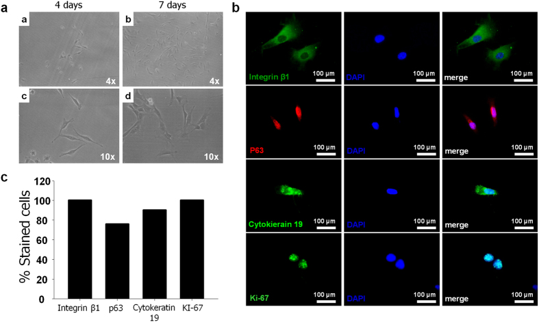
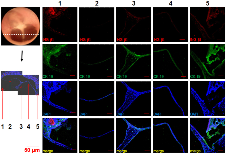
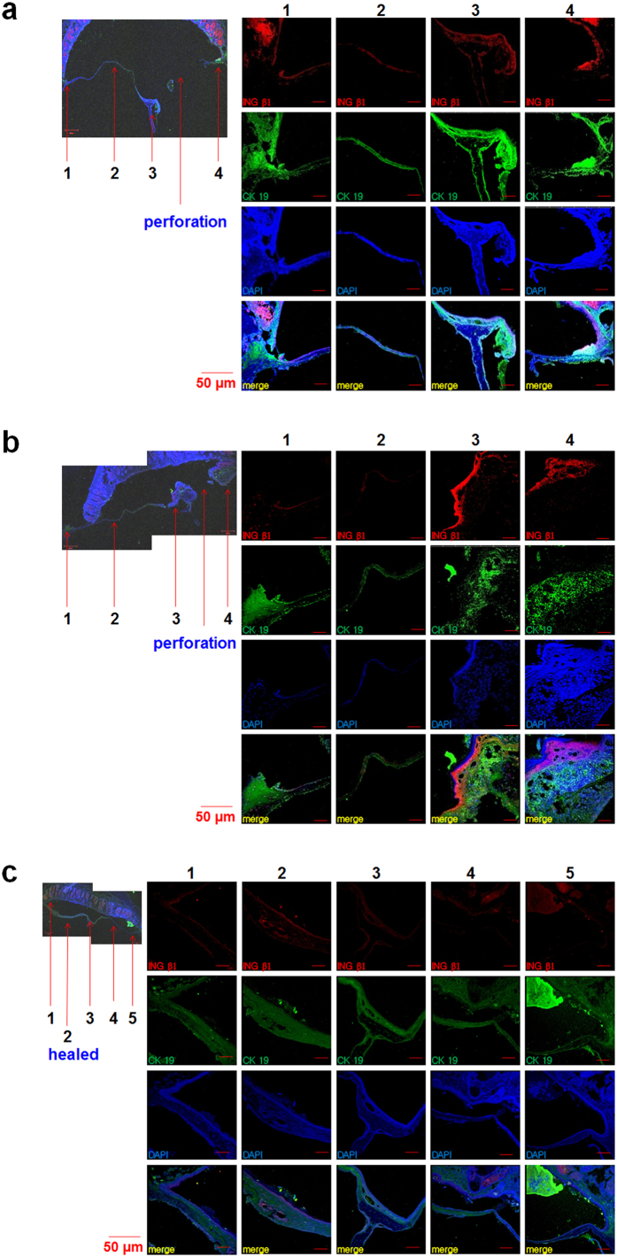
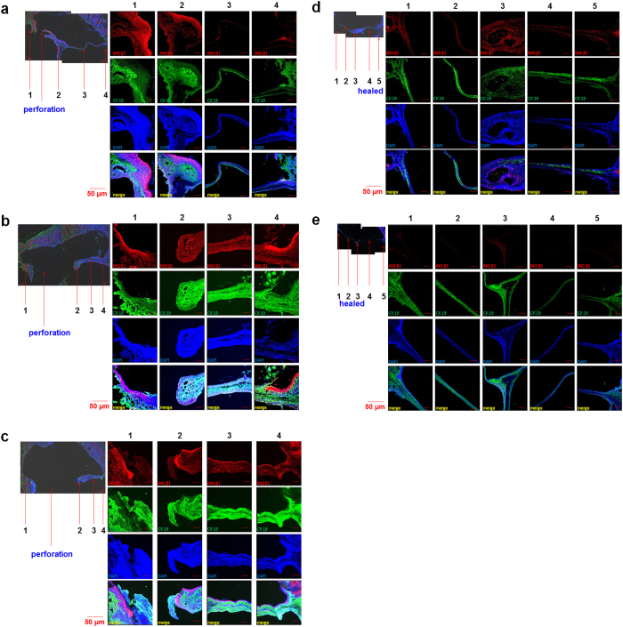
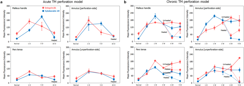
Tympanic membrane (TM) perforation, in particular chronic otitis media, is one of the most common clinical problems in the world and can present with sensorineural healing loss. Here, we explored an approach for TM regeneration where the latent progenitor or stem cells within TM epithelial layers may play an important regulatory role. We showed that potential TM stem cells present highly positive staining for epithelial stem cell markers in all areas of normal TM tissue. Additionally, they are present at high levels in perforated TMs, especially in proximity to the holes, regardless of acute or chronic status, suggesting that TM stem cells may be a potential factor for TM regeneration. Our study suggests that latent TM stem cells could be potential regulators of regeneration, which provides a new insight into this clinically important process and a potential target for new therapies for chronic otitis media and other eardrum injuries.
Figures
Similar articles
-
Tympanic Membrane Derived Stem Cell-Like Cultures for Tissue Regeneration.Stem Cells Dev. 2018 May 15;27(10):649-657. doi: 10.1089/scd.2018.0021. Epub 2018 Apr 23. Stem Cells Dev. 2018. PMID: 29571277
-
Stem cells and enhanced healing of chronic tympanic membrane perforation.Acta Otolaryngol. 2008 Apr;128(4):352-9. doi: 10.1080/00016480701762508. Acta Otolaryngol. 2008. PMID: 18368564
-
Tympanic membrane regeneration using a water-soluble chitosan patch.Tissue Eng Part A. 2010 Jan;16(1):225-32. doi: 10.1089/ten.TEA.2009.0476. Tissue Eng Part A. 2010. PMID: 19691425
-
3D printing technology and applied materials in eardrum regeneration.J Biomater Sci Polym Ed. 2023 May;34(7):950-985. doi: 10.1080/09205063.2022.2147350. Epub 2022 Nov 24. J Biomater Sci Polym Ed. 2023. PMID: 36373498 Review.
-
Can Tissue Engineering Bring Hope to the Development of Human Tympanic Membrane?Tissue Eng Part B Rev. 2021 Dec;27(6):572-589. doi: 10.1089/ten.TEB.2020.0176. Epub 2021 Feb 17. Tissue Eng Part B Rev. 2021. PMID: 33164696 Review.
Cited by
-
A Hierarchy of Proliferative and Migratory Keratinocytes Maintains the Tympanic Membrane.Cell Stem Cell. 2021 Feb 4;28(2):315-330.e5. doi: 10.1016/j.stem.2020.10.006. Epub 2020 Nov 11. Cell Stem Cell. 2021. PMID: 33181078 Free PMC article.
-
Mimicking the Human Tympanic Membrane: The Significance of Scaffold Geometry.Adv Healthc Mater. 2021 Jun;10(11):e2002082. doi: 10.1002/adhm.202002082. Epub 2021 May 4. Adv Healthc Mater. 2021. PMID: 33945239 Free PMC article.
-
Stem Cell-Induced Inflammation in Cholesteatoma is Inhibited by the TLR4 Antagonist LPS-RS.Cells. 2020 Jan 14;9(1):199. doi: 10.3390/cells9010199. Cells. 2020. PMID: 31947538 Free PMC article.
-
Stem cells in middle ear cholesteatoma contribute to its pathogenesis.Sci Rep. 2018 Apr 18;8(1):6204. doi: 10.1038/s41598-018-24616-4. Sci Rep. 2018. PMID: 29670222 Free PMC article.
-
Inhibition of TGF-β signaling enables long-term proliferation of mouse primary epithelial stem/progenitor cells of the tympanic membrane and the middle ear mucosa.Sci Rep. 2023 Mar 20;13(1):4532. doi: 10.1038/s41598-023-31246-y. Sci Rep. 2023. PMID: 36941290 Free PMC article.
References
-
- Gladstone H. B., Jackler R. K. & Varav K. Tympanic membrane wound healing. An overview. Otolaryngologic clinics of North America 28, 913–932 (1995). - PubMed
-
- Wang W. Q., Wang Z. M. & Chi F. L. Spontaneous healing of various tympanic membrane perforations in the rat. Acta oto-laryngologica 124, 1141–1144 (2004). - PubMed
-
- Laidlaw D. W. et al. Tympanic membrane repair with a dermal allograft. The Laryngoscope 111, 702–707 (2001). - PubMed
-
- Kim J. H. et al. Tympanic membrane regeneration using a water-soluble chitosan patch. Tissue Engineering Part A 16, 225–232 (2009). - PubMed
-
- Kim J. et al. A healing method of tympanic membrane perforations using three-dimensional porous chitosan scaffolds. Tissue Engineering Part A 17, 2763–2772 (2011). - PubMed
Publication types
MeSH terms
Substances
LinkOut - more resources
Full Text Sources
Other Literature Sources
Medical
