. 2015 Oct;3(10):1185-92.
doi: 10.1158/2326-6066.CIR-15-0102. Epub 2015 Jun 22.

Radiographic Profiling of Immune-Related Adverse Events in Advanced Melanoma Patients Treated with Ipilimumab

Affiliations

Radiographic Profiling of Immune-Related Adverse Events in Advanced Melanoma Patients Treated with Ipilimumab

Sree Harsha Tirumani et al. Cancer Immunol Res. 2015 Oct.

Abstract

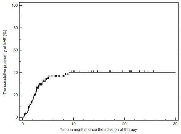
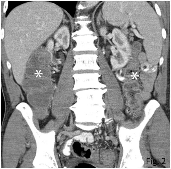
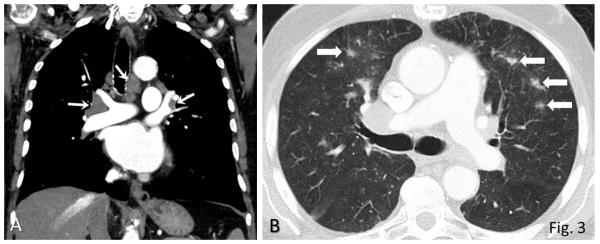
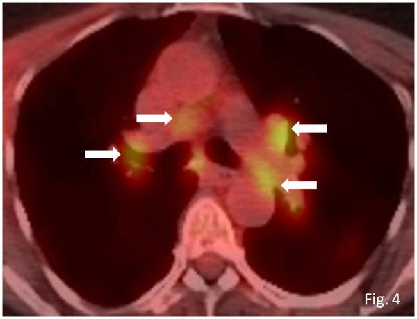
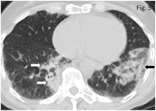
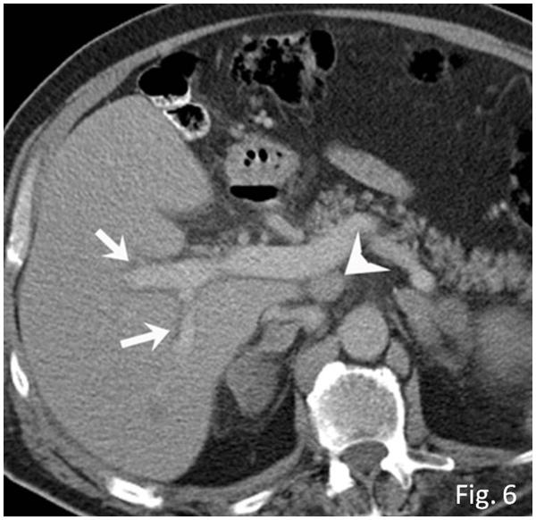
Ipilimumab is a promising novel immunotherapy agent and is associated with a variety of immune-related adverse events (irAE). The purpose of this study was to investigate the manifestations of irAEs on body imaging in patients with advanced melanoma treated with ipilimumab. One-hundred forty-seven patients with advanced melanoma (59 women, 88 men; median age, 64.5 years) treated with ipilimumab were studied. All patients had the baseline and at least one follow-up chest/abdomen/pelvis CT or PET/CT during therapy, which were reviewed by a consensus of two radiologists blinded to the clinical data. Findings indicative of individual types of irAEs were assessed, including thyroiditis, sarcoid-like lymphadenopathy, pneumonitis, hepatitis, pancreatitis, and colitis. Among the 147 patients, 46 (31%) had radiologically identified irAEs. The time interval from the initiation of therapy to the development of irAEs was less than 3 months in 76% (35 of 46) of the patients (range, 0.2-9.1 months). Clinical characteristics did not differ between patients with and without irAEs (P > 0.18). Among the individual types of irAEs, colitis was most common (n = 28; 19%), followed by sarcoid-like lymphadenopathy (n = 8; 5%) and pneumonitis (n = 8; 5%). Hepatitis (n = 3), thyroiditis (n = 2), and pancreatitis (n = 1) were less common. The resolution of irAEs was noted in 32 of 36 patients (89%) with further follow-up scans, with a median time of 2.3 months after the detection of irAE. In conclusion, irAEs were noted on body imaging in 31% of patients with melanoma treated with ipilimumab. Colitis was the most common, followed by sarcoid-like lymphadenopathy and pneumonitis. The results call for an increased awareness of irAEs, given the expanding role of cancer immunotherapy.


Figures

Fig. 1
Fig. 1
The cumulative probability of radiographically-evident immune-related adverse events (irAEs) during ipilimumab therapy.
Fig. 2
Fig. 2
Colitis with a diffuse colitis pattern in a 64-year-old man with advanced melanoma treated with ipilimumab, presenting with diarrhea. Coronal reformatted contrast-enhanced CT image of the abdomen obtained at 2.6 months after the initiation of ipilimumab treatment demonstrated a new fluid-filled dilated colon (asterisk) with mucosal hyperemia indicating diffuse colitis. Colonoscopic biopsy confirmed colonic inflammation with mucosal injury consistent with ipilimumab-associated colitis. Patient was treated with oral steroids followed by intravenous infliximab, leading to resolution of the findings on the follow-up scan at 1.8 months after the onset (data not shown).
Fig. 3
Fig. 3
Sarcoid-like lymphadenopathy in an asymptomatic 81-year-old man with metastatic melanoma treated with ipilimumab. A. Coronal reformatted contrast-enhanced chest CT performed at 4.9 months after the initiation of ipilimumab therapy demonstrated new bilateral symmetric mediastinal and hilar lymphadenopathy, resembling sarcoidosis. B. Axial CT image of the lungs showed bilateral irregular and nodular parenchymal opacities in upper and middle lung predominance (arrows), with peribronchovascular involvement, which falls in the spectrum of lung parenchymal manifestations of pulmonary sarcoidosis.
Fig. 4
Fig. 4
Sarcoid-like lymphadenopathy in an asymptomatic 55-year-old woman with metastatic melanoma treated with ipilimumab. Axial fused FDG-PET/CT images at 3 months of ipilimumab therapy demonstrate new FDG-avid mediastinal and bilateral hilar lymphadenopathy (arrows) mimicking sarcoidosis. A follow-up PET/CT performed 5 months later showed resolution of FDG-avid lymphadenopathy (data not shown).
Fig. 5
Fig. 5
Pneumonitis in a 72-year-old woman with metastatic melanoma treated with ipilimumab. Axial CT image of the chest obtained at 4.0 months after the initiation of ipilimumab therapy demonstrates new bilateral consolidative and ground glass opacities predominantly in peripheral and lower distribution (arrows), mimicking cryptogenic organizing pneumonia (COP) pattern. Patient was symptomatic with cough at this time and was treated with oral steroids. Further follow-up CT scan performed 2.1 months after the onset demonstrated resolution of the findings (data not shown).
Fig. 6
Fig. 6
Hepatitis in a 63-year-old man with metastatic melanoma treated with ipilimumab. Axial contrast-enhanced CT scan of the abdomen performed at 2.7 months of ipilimumab therapy demonstrates new periportal edema (arrows) with hepatomegaly and new periportal lymphadenopathy (arrowhead). Markedly elevated liver functions were noted, leading to liver biopsy, which revealed severe panlobular hepatitis with lymphoplasmacytic infiltrate and eosinophils, and foci of central vein damage and perivenular collapse, consistent with ipilimumab-associated hepatitis. The patient was treated with steroids and mycophenolate, and liver functions were normalized.

References

    1. Hodi FS, O'Day SJ, McDermott DF, Weber RW, Sosman JA, Haanen JB, et al. Improved survival with ipilimumab in patients with metastatic melanoma. N Engl J Med. 2010;363:711–23. - PMC - PubMed
    1. Leach DR, Krummel MF, Allison JP. Enhancement of antitumor immunity by CTLA-4 blockade. Science. 1996;271:1734–6. - PubMed
    1. Brunet JF, Denizot F, Luciani MF, Roux-Dosseto M, Suzan M, Mattei MG, et al. A new member of the immunoglobulin superfamily--CTLA-4. Nature. 1987;328:267–70. - PubMed
    1. Freeman GJ, Gribben JG, Boussiotis VA, Ng JW, Restivo VA, Jr, Lombard LA, et al. Cloning of B7-2: a CTLA-4 counter-receptor that costimulates human T cell proliferation. Science. 1993;262:909–11. - PubMed
    1. Hodi FS, Mihm MC, Soiffer RJ, Haluska FG, Butler M, Seiden MV, et al. Biologic activity of cytotoxic T lymphocyte-associated antigen 4 antibody blockade in previously vaccinated metastatic melanoma and ovarian carcinoma patients. Proc Natl Acad Sci U S A. 2003;100:4712–7. - PMC - PubMed

MeSH terms