

From G Protein-coupled Receptor Structure Resolution to Rational Drug Design
- PMID: 26100628
- PMCID: PMC4528114
- DOI: 10.1074/jbc.R115.668251
From G Protein-coupled Receptor Structure Resolution to Rational Drug Design
Abstract
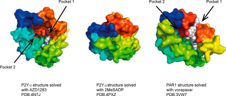
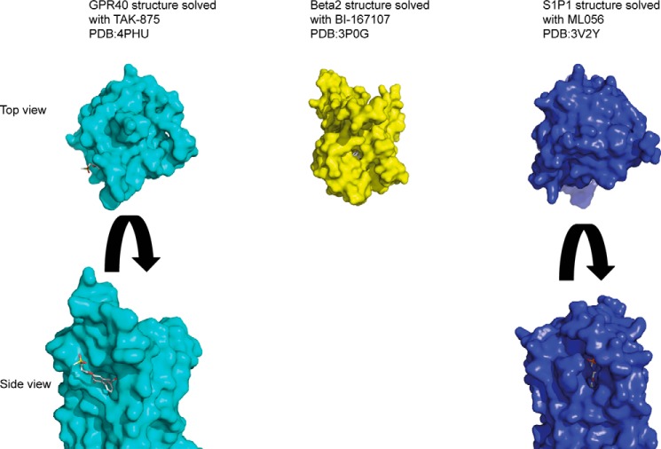
A number of recent technical solutions have led to significant advances in G protein-coupled receptor (GPCR) structural biology. Apart from a detailed mechanistic view of receptor activation, the new structures have revealed novel ligand binding sites. Together, these insights provide avenues for rational drug design to modulate the activities of these important drug targets. The application of structural data to GPCR drug discovery ushers in an exciting era with the potential to improve existing drugs and discover new ones. In this review, we focus on technical solutions that have accelerated GPCR crystallography as well as some of the salient findings from structures that are relevant to drug discovery. Finally, we outline some of the approaches used in GPCR structure based drug design.
Keywords: G protein-coupled receptor (GPCR); X-ray crystallography; drug design; drug discovery; membrane protein.
© 2015 by The American Society for Biochemistry and Molecular Biology, Inc.
Figures
Similar articles
-
Novel Allosteric Modulators of G Protein-coupled Receptors.J Biol Chem. 2015 Aug 7;290(32):19478-88. doi: 10.1074/jbc.R115.662759. Epub 2015 Jun 22. J Biol Chem. 2015. PMID: 26100627 Free PMC article. Review.
-
Thematic Minireview Series: New Directions in G Protein-coupled Receptor Pharmacology.J Biol Chem. 2015 Aug 7;290(32):19469-70. doi: 10.1074/jbc.R115.675728. Epub 2015 Jun 30. J Biol Chem. 2015. PMID: 26126823 Free PMC article. Review.
-
GPCR structures in drug design, emerging opportunities with new structures.Bioorg Med Chem Lett. 2014 Sep 1;24(17):4073-9. doi: 10.1016/j.bmcl.2014.07.009. Epub 2014 Jul 10. Bioorg Med Chem Lett. 2014. PMID: 25086683 Review.
-
Allosteric inhibition of g-protein coupled receptor oligomerization: strategies and challenges for drug development.Curr Top Med Chem. 2014;14(15):1842-63. doi: 10.2174/1568026614666140901130843. Curr Top Med Chem. 2014. PMID: 25175995 Review.
-
Small Molecule Allosteric Modulators of G-Protein-Coupled Receptors: Drug-Target Interactions.J Med Chem. 2019 Jan 10;62(1):24-45. doi: 10.1021/acs.jmedchem.7b01844. Epub 2018 Feb 26. J Med Chem. 2019. PMID: 29457894
Cited by
-
Emerging Computational Methods for the Rational Discovery of Allosteric Drugs.Chem Rev. 2016 Jun 8;116(11):6370-90. doi: 10.1021/acs.chemrev.5b00631. Epub 2016 Apr 13. Chem Rev. 2016. PMID: 27074285 Free PMC article. Review.
-
Supporting the Identification of Novel Fragment-Based Positive Allosteric Modulators Using a Supervised Molecular Dynamics Approach: A Retrospective Analysis Considering the Human A2A Adenosine Receptor as a Key Example.Molecules. 2017 May 16;22(5):818. doi: 10.3390/molecules22050818. Molecules. 2017. PMID: 28509867 Free PMC article.
-
Rapid and accurate assessment of GPCR-ligand interactions Using the fragment molecular orbital-based density-functional tight-binding method.J Comput Chem. 2017 Sep 5;38(23):1987-1990. doi: 10.1002/jcc.24850. Epub 2017 Jul 4. J Comput Chem. 2017. PMID: 28675443 Free PMC article.
-
Molecular docking as a popular tool in drug design, an in silico travel.Adv Appl Bioinform Chem. 2016 Jun 28;9:1-11. doi: 10.2147/AABC.S105289. eCollection 2016. Adv Appl Bioinform Chem. 2016. PMID: 27390530 Free PMC article. Review.
-
Human Adenosine A2A Receptor: Molecular Mechanism of Ligand Binding and Activation.Front Pharmacol. 2017 Dec 14;8:898. doi: 10.3389/fphar.2017.00898. eCollection 2017. Front Pharmacol. 2017. PMID: 29311917 Free PMC article. Review.
References
-
- Paul S. M., Mytelka D. S., Dunwiddie C. T., Persinger C. C., Munos B. H., Lindborg S. R., Schacht A. L. (2010) How to improve R&D productivity: the pharmaceutical industry's grand challenge. Nat. Rev. Drug Discov. 9, 203–214 - PubMed
-
- Erlanson D. A. (2012) Introduction to fragment-based drug discovery. Top. Curr. Chem. 317, 1–32 - PubMed
-
- Schindler T., Bornmann W., Pellicena P., Miller W. T., Clarkson B., Kuriyan J. (2000) Structural mechanism for STI-571 inhibition of Abelson tyrosine kinase. Science 289, 1938–1942 - PubMed
Publication types
MeSH terms
Substances
LinkOut - more resources
Full Text Sources
