Review
. 2015 Jul;11(7):401-13.
doi: 10.1038/nrneurol.2015.99. Epub 2015 Jun 23.

Interactions of the histamine and hypocretin systems in CNS disorders

Affiliations
Review

Interactions of the histamine and hypocretin systems in CNS disorders

Ling Shan et al. Nat Rev Neurol. 2015 Jul.

Abstract

Histamine and hypocretin neurons are localized to the hypothalamus, a brain area critical to autonomic function and sleep. Narcolepsy type 1, also known as narcolepsy with cataplexy, is a neurological disorder characterized by excessive daytime sleepiness, impaired night-time sleep, cataplexy, sleep paralysis and short latency to rapid eye movement (REM) sleep after sleep onset. In narcolepsy, 90% of hypocretin neurons are lost; in addition, two groups reported in 2014 that the number of histamine neurons is increased by 64% or more in human patients with narcolepsy, suggesting involvement of histamine in the aetiology of this disorder. Here, we review the role of the histamine and hypocretin systems in sleep-wake modulation. Furthermore, we summarize the neuropathological changes to these two systems in narcolepsy and discuss the possibility that narcolepsy-associated histamine abnormalities could mediate or result from the same processes that cause the hypocretin cell loss. We also review the changes in the hypocretin and histamine systems, and the associated sleep disruptions, in Parkinson disease, Alzheimer disease, Huntington disease and Tourette syndrome. Finally, we discuss novel therapeutic approaches for manipulation of the histamine system.


Figures

Figure 1 |
Figure 1 |
Neurons containing histamine, hypocretin and melanin-concentrating hormone in the human hypothalamus. a | Overview of the medial surface of the human brain, and b | detail of the hypothalamus. The distribution of neurons containing histamine, hypocretin and melanin-concentrating hormone are shown with in situ hybridization in coronal sections of hypothalamus at the c | posterior level through the medial mammillary nucleus, d | rostromammillary level at the principal mammillary fasciculus, e | level of the caudal end of the fornix and f | premamillary level. In panels c–f, the third ventricle is on the left side. Abbreviations: F, fornix; HCRT, hypocretin neurons; HDC, histidine-decarboxylase-positive (histamine) neurons; LHA, lateral hypothalamic area; LTu, lateral segment of the lateral tuberal nucleus; MCH, melanin-concentrating hormone neurons; MM, medial mammillary nucleus; MP, mammillary peduncle; MT, mammillothalamic tract; TMc, caudal tuberomammillary nucleus; TMl; lateral tuberomammillary nucleus; TMm, medial tuberomammillary nucleus; TMv, ventral tuberomammillary nucleus. Panels a and b reprinted with permission from Elsevier © Handbook of Clinical Neurology Vol. 79. Swaab, D. F. The human hypothalamus: basic and clinical aspects. Part 1: Nuclei of the human hypothalamus 3–38 (2003). Parts c–f adapted with permission from John Wiley & Sons © Krolewski, D. M. et al. J. Comp. Neurol. 518, 4591–4611 (2010).
Figure 2 |
Figure 2 |
The hypocretin and histamine systems in the mouse brain—origin and general projections. Red dots indicate the location and projections of hypocretin neurons. The blue dots and lines represent the location and innervations of histamine neurons. Mouse brain connectivity was mapped using hypocretin–Cre and histidine-decarboxylase–Cre mouse lines. Adeno-associated viral vectors expressing Cre-dependent enhanced green fluorescent protein was injected to trace axonal projections from both hypocretin (red) and histamine (blue) neurons. Abbreviations: HCRT, hypocretin neurons; HDC, histidine-decarboxylase-positive (histamine) neurons. Reprinted with permission. © 2014 Allen Institute for Brain Science. Allen Mouse Brain Connectivity Atlas [online]. Available from: http://connectivity.brain-map.org/.
Figure 3 |
Figure 3 |
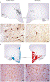
Loss of hypocretin neurons and increase in histamine neurons in narcolepsy. a | Number and distribution of hypocretin neurons, and a representative cell staining of brain slices from a healthy control (left) and an individual with narcolepsy (right). Scale bars = 25 μm. b | Number and distribution of histamine neurons, and a representative cell staining from a healthy control (left) and an individual with narcolepsy (right). Scale bars = 50 μm. Part a reprinted with permission from Elsevier © Thannickal, T. C. et al. Reduced number of hypocretin neurons in human narcolepsy. Neuron 27, 469–474 (2000). Part b adapted with permission from John Wiley & Sons © John, J. et al. Ann. Neurol. 74, 786–793 (2013).
Figure 4 |
Figure 4 |
A schematic model of autoimmune-triggered histaminergic involvement in hypocretin neuron degeneration. a | The development of narcolepsy can sometimes be triggered by environmental factors, such as Streptococcus pyogenes infection, upper airway infection, H1N1 influenza or H1N1 vaccination with squalene-based adjuvant. Histaminergic signalling has a crucial role in several autoimmune diseases of the CNS. b | For example, binding of histamine to endothelial cell histamine receptors increases the permeability of the blood–brain barrier, (as indicated by increased levels of neuron-specific markers Aβ, total tau protein, phosphorylated tau, and neuro-specific enolase in the CSF), thereby facilitating T cell entry to the CNS. Moreover, histamine signalling enhances the activity of type 1 T helper cells through binding to histamine type 1 receptors located on these cells. c | T cells, B cells, macrophages, microglia and mast cells secrete histamine and other cytokines and chemokines,,,, triggering a local inflammatory response that can damage the sensitive hypocretin neurons. Abbreviations: Aβ, amyloid-β; CSF, cerebrospinal fluid.
Figure 5 |
Figure 5 |
Specific degeneration of histamine, MCH and hypocretin neurons in PD. The course of PD can be divided into several stages, including unaffected, preclinical PD, and PD (from left to right). a | The number of histamine neurons in the TMN remains stable despite the accumulation of Lewy bodies and Lewy neurites. TMN neurons were stained for α-synuclein (brown) and counterstained by thionin (blue). Scale bar = 250 μm. The numbers of b | MCH neurons and c | hypocretin neurons decrease in line with increasing disease severity. MCH and hypocretin neurons were immunostained by their peptide-specific antibodies, followed by the diaminobenzidine colouring (reddish brown). Scale bars = 50 μm. Abbreviations: MCH, melanin-concentrating hormone; PD, Parkinson disease; TMN, tuberomamillary nucleus. Part a reprinted with permission from Elsevier Ltd © Shan, L. et al. Neuronal histamine production remains unaltered in Parkinson’s disease despite the accumulation of Lewy bodies and Lewy neurites in the tuberomamillary nucleus. Neurobiol. Aging 33, 1343–1344 (2012). Parts b and c adapted with permission from Oxford University Press © Thannickal, T. C. et al. Hypocretin (orexin) cell loss in Parkinson’s disease. Brain 130, 1586–1595 (2007).
Figure 6 |
Figure 6 |
Effects of pitolisant, modafinil and placebo on patients with narcolepsy. Changes in Epworth sleepiness scale (ESS) score from the beginning of a double-blind, randomized phase III study (‘Baseline’) to the end of the 9-week treatment period (‘Final’). Data points indicate the mean ESS score; error bars indicate SEM. *A significant reduction in ESS was found for both modafinil and pitolisant versus placebo. No significant difference in ESS was found between pitolisant and modafinil.

Similar articles

Cited by

References

    1. von Economo C Sleep as a problem of localization. J. Nerv. Ment. Dis 71, 249–259 (1930).
    1. Peyron C et al. A mutation in a case of early onset narcolepsy and a generalized absence of hypocretin peptides in human narcoleptic brains. Nat. Med 6, 991–997 (2000). - PubMed
    1. Thannickal TC et al. Reduced number of hypocretin neurons in human narcolepsy. Neuron 27, 469–474 (2000). - PMC - PubMed
    1. American Academy of Sleep Medicine. International Classification of Sleep Disorders: Diagnostic and Coding Manual 3rd edn (AASM, 2014).
    1. John J et al. Greatly increased numbers of histamine cells in human narcolepsy with cataplexy. Ann. Neurol 74, 786–793 (2013). - PMC - PubMed

Publication types

MeSH terms