. 2015 Jul 7;112(27):E3609-18.
doi: 10.1073/pnas.1507704112. Epub 2015 Jun 22.

Allosteric interactions between agonists and antagonists within the adenosine A2A receptor-dopamine D2 receptor heterotetramer

Affiliations

Allosteric interactions between agonists and antagonists within the adenosine A2A receptor-dopamine D2 receptor heterotetramer

Jordi Bonaventura et al. Proc Natl Acad Sci U S A. .

Abstract

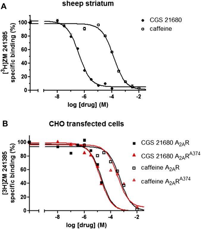
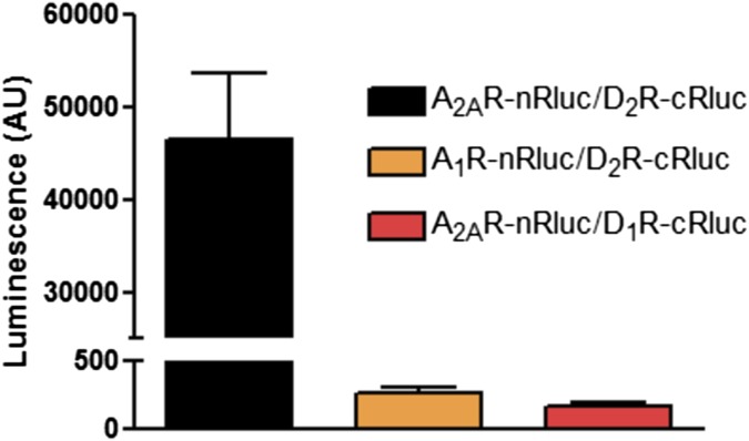
Adenosine A2A receptor (A2AR)-dopamine D2 receptor (D2R) heteromers are key modulators of striatal neuronal function. It has been suggested that the psychostimulant effects of caffeine depend on its ability to block an allosteric modulation within the A2AR-D2R heteromer, by which adenosine decreases the affinity and intrinsic efficacy of dopamine at the D2R. We describe novel unsuspected allosteric mechanisms within the heteromer by which not only A2AR agonists, but also A2AR antagonists, decrease the affinity and intrinsic efficacy of D2R agonists and the affinity of D2R antagonists. Strikingly, these allosteric modulations disappear on agonist and antagonist coadministration. This can be explained by a model that considers A2AR-D2R heteromers as heterotetramers, constituted by A2AR and D2R homodimers, as demonstrated by experiments with bioluminescence resonance energy transfer and bimolecular fluorescence and bioluminescence complementation. As predicted by the model, high concentrations of A2AR antagonists behaved as A2AR agonists and decreased D2R function in the brain.

Keywords: GPCR heteromers; adenosine A2A receptor; caffeine; dopamine D2 receptor.


Conflict of interest statement

The authors declare no conflict of interest.

Figures

Fig. 1.
Fig. 1.
Effect of an A2AR agonist and caffeine on [3H]quinpirole binding to D2R. [3H]Quinpirole binding (6 nM) was determined in membrane preparations from sheep striatum (black bars) or CHO cells transfected with D2R cDNA (2 µg) and A2AR cDNA (3 µg) (red bars) or D2R cDNA (2 µg) and cDNA (3 µg) from mutated A2AR (A2AA374R; blue bars) in the presence or the absence of increasing concentrations of the A2AR agonist CGS21680 (A) or caffeine (B). Values are mean ± SEM from between three and five different experiments of relative [3H]quinpirole-specific binding (% of nontreated membranes). Statistical significance was calculated by one-way ANOVA followed by Dunnett’s post hoc test. *P < 0.05; **P < 0.01, compared with nontreated membrane preparations.
Fig. S1.
Fig. S1.
Competition curves of [3H]ZM 241385 vs. CGS 21680 or caffeine. Membrane preparations from sheep striatum (A) or CHO cells (B) transfected with D2R cDNA (2 µg) and A2AR cDNA (3 µg) (B, black curves) or with D2R cDNA (2 µg) and A2AA374R cDNA (or 3 µg) (B, red curves) were incubated with [3H]ZM 241385 (2 nM) and increasing concentrations of CGS 21680 (10 nM–10 µM) or caffeine (1 µM–10 mM). Values are mean ± SEM from a representative experiment performed in triplicate of the relative [3H]ZM 241385-specific binding, where 100% corresponds to 0.96 ± 0.04 pmol/mg protein for sheep striatum and to 0.74 ± 0.03 pmol/mg protein for CHO cells transfected with A2AR or 0.90 ± 0.03 for CHO cells transfected with A2AA374R.
Fig. 2.
Fig. 2.
Biphasic effect of caffeine and selective A2AR antagonists on [3H]quinpirole binding and D2R-mediated ERK1/2 phosphorylation. (A, C, and E) [3H]Quinpirole binding (6 nM) was determined in membrane preparations from sheep striatum not preincubated (control, blue bars) or preincubated (black bars) for 30 min with the A2AR agonist CGS 21680 (100 nM) and increasing concentrations of caffeine (A) or the selective A2AR antagonists SCH 58216 (C) or KW 6002 (E). Values are mean ± SEM from four to eight different experiments of relative [3H]quinpirole binding (% of nontreated control membranes, c). Statistical significance was calculated by one-way ANOVA followed by the Newman–Keuls post hoc test. *P < 0.05; **P < 0.01; ***P < 0.001, compared with c. #P < 0.05; ##P < 0.01; ###P < 0.001 compared with only CGS 21680. (B, D, and F) ERK1/2 phosphorylation was determined in HEK-293 cells transfected with D2R cDNA (0.8 µg) and A2AR cDNA (1.2 µg), stimulated for 5 min with CGS 21680 (CGS; 100 nM) or quinpirole (QP; 1 µM) alone (orange and blue bars, respectively) or in combination (black bars) after incubation for 10 min with vehicle or with caffeine (B), SCH 58126 (D), or KW 6002 (F). ERK1/2 phosphorylation was quantified; values represent mean ± SEM from three to six different experiments of the percentage of phosphorylation relative to basal levels in nontreated cells (100%). Statistical significance was calculated by one-way ANOVA followed by Dunnett’s post hoc test. *P < 0.05; **P < 0.01; ***P < 0.001, compared with QP. #P < 0.05; ##P < 0.01; ###P < 0.001, compared with cells treated with QP plus CGS.
Fig. 3.
Fig. 3.
Dissociation kinetics of [3H]ZM 241385 in the presence of caffeine or selective A2AR ligands. Dissociation curves of the A2AR antagonist [3H]ZM 241385 (1.5 nM) in the absence (black curve) or presence of either the A2AR antagonists SCH 58260 (10 nM, blue curve) or caffeine (30 µM, green curve), or the A2AR agonist CGS 21680 (10 nM, red curve). Data points are means ± SD of triplicates. Fitted Koff values of [3H]ZM 241385 dissociation were 0.025 ± 0.002 min−1 (i.e., a residence time of 40 min) for control, 0.025 ± 0.003 min−1 (residence time of 40 min) in the presence of SCH 58260, and 0.028 ± 0.004 min−1 (residence time of 36 min) in the presence of caffeine. A biphasic curve was obtained in the presence of CGS 21680 (red curve) with a Koff1 value of 0.19 ± 0.03 min−1 and a Koff2 value of 0.004 ± 0.003 min−1 (residence time of 5 and 250 min, respectively).
Fig. S2.
Fig. S2.
Bioluminescence complementation within the A2AR-D2R heteromer. Luminescence due to Rluc in HEK-293 cells cotransfected with A2AR-nRluc and D2R-cRluc, A1R-nRluc and D2R-cRluc, or A2AR-nRluc and D1R-cRluc. On addition of coelenterazine H, strong luminescence could be observed only in cells coexpressing A2AR-nRluc and D2R-cRluc.
Fig. 4.
Fig. 4.
Tetrameric structure of the A2AR-D2R heteromer. (A) Fluorescence due to complementation [in arbitrary units (AU)] of YFP Venus was determined in HEK-293 cells coexpressing A2AR-nYFP and A2AR-cYFP, D2R-nYFP and D2R-cYFP, or A2AR-nYFP and D2R-cYFP either not treated or treated with the indicated HIV TAT peptides (4 µM) for 4 h. Values represent means ± SEM from seven or eight different experiments. Statistical significance was calculated by one-way ANOVA followed by Dunnett’s post hoc test. **P < 0.01, compared with the nontreated cells. (B) BRET was determined in cells expressing A2AR-nRluc, D2R-cRluc, A2AR-nYFP and D2R-cYFP, or A2AR-nYFP and D2R-cYFP and the respective controls replacing A2AR for A1R or D2R for D1R. Values are mean ± SEM of three different experiments. (Upper) Schematic representation of BRET with bimolecular luminescence and fluorescence complementation.
Fig. 5.
Fig. 5.
Allosteric modulation of A2AR antagonists on D2R-mediated modulation of neuronal function. (A and B) Effect of the A2R antagonist SCH 58261 on NMDA-mediated depolarized plateau potential on D2R-responsive neurons in rat ventral striatal slices. (A) Consecutive traces showing typical transitions where the action of NMDA (5 µM) was recorded before and in the presence of D2R NPA (10 µM) and the A2AR antagonist SCH 58261 (1 µM). On a D2R-responsive neuron, subsequent application of SCH 58261 totally counteracts the effect of NPA, i.e., inhibition of the depolarized plateau potential and firing frequency. (B) Summary histogram obtained from D2R-responsive neurons illustrating the antagonistic effect of SCH 58261 on the action potential firing frequency. Data represent mean ± SEM (n = 7). Statistical significance was calculated by one-way ANOVA followed by Dunnett’s post hoc test. ***P < 0.001, compared with the untreated slice preparation (c). (C) Locomotor activity in nonhabituated rats during the first 20 min after the administration of vehicle or the A2AR antagonist KW 6002 (1–30 mg/kg, i.p.). The A2AR agonist CGS 21680 (0.1 mg/kg i.p.), or vehicle, was administered 30 min before the administration of KW 6002. A high dose of KW 6002 produced significant locomotor depression, which was counteracted by a previous administration of the additional depressant dose of CGS 21680. Statistical significance was calculated by one-way ANOVA followed by Dunnett’s post hoc test. **P < 0.01 compared with controls (animals only treated with vehicle).
Fig. S3.
Fig. S3.
Locomotor depressant effects of high doses of caffeine. Locomotor activity in nonhabituated rats during the first 20 min after the administration of vehicle or caffeine (30–100 mg/kg, i.p.). The A2AR agonist CGS 21680 (0.1 mg/kg i.p.) or vehicle was administered 30 min before the administration of caffeine. High doses of caffeine produced significant locomotor depression, which was neither counteracted nor potentiated by a previous administration of the additional depressant dose of CGS 21680. Statistical significance was calculated by one-way ANOVA followed by Dunnett’s post hoc test. *P < 0.05; **P < 0.01, compared with controls (animals treated only with vehicle).
Fig. 6.
Fig. 6.
Effect of an A2AR agonist and caffeine on [3H]raclopride binding. (A and B) [3H]Raclopride (4 nM) binding was determined in membrane preparations from sheep striatum (black bars), human caudate nucleus (white bars), or CHO cells transfected with D2R cDNA (2 µg) and A2AR cDNA (3 µg; red bars), D2R cDNA (2 µg) and cDNA (3 µg) from mutated A2AR (A2AA374R; blue bars), or CHO cells transfected only with D2R cDNA (2 µg; green bars) in the presence or the absence of increasing concentrations of the A2AR agonist CGS21680 (A) or caffeine (B). (C) [3H]raclopride (4 nM) binding determined in membrane preparations from sheep striatum either untreated (white bar, c) or treated with CGS 21680 (10 µM) in the absence or presence of increasing concentrations of caffeine (black bars). Values are mean ± SEM from three to five different experiments) of the relative [3H]raclopride-specific binding (% of nontreated membranes). Statistical significance was calculated by one-way ANOVA followed by Dunnett’s post hoc test or the Newman–Keuls post hoc test. *P < 0.05; **P < 0.01; ***P < 0.001, compared with the untreated membrane preparations. #P < 0.05; ##P < 0.01, compared with the membranes treated only with CGS 21680.
Fig. S4.
Fig. S4.
Effect of A1R agonist or antagonist on [3H]raclopride binding. (A and B) [3H]Raclopride binding (4 nM) was determined in membrane preparations from sheep striatum (black bars) or human caudate nucleus (red bars) in the presence or the absence of increasing concentrations of the A1R agonist CCPA (A) or the antagonist DPCPX (B), with higher selectivity for A1R over A2AR. Values are means ± SEM (three to five different experiments) of the relative [3H]raclopride-specific binding (% respect to samples without A1R ligands). Statistical significance was calculated by one-way ANOVA, followed by Dunnett’s post hoc test. *P < 0.05; **P < 0.01, compared with the untreated membrane preparations. (C) Competition curve of the A2AR antagonist [3H]ZM 241385 (2 nM) vs. the A1R-selective antagonist DPCPX. Membrane preparations from sheep striatum were incubated with [3H]ZM 241385 (2 nM) and increasing concentrations of DPCPX (1 nM–300 µM). Values are mean ± SEM from a representative experiment performed in triplicate of the relative [3H]ZM 241385-specific binding, where 100% corresponds to 0.96 ± 0.05 pmol/mg protein.
Fig. 7.
Fig. 7.
Detection of A2AR-D2R heteromers in sheep striatum and effect of HIV TAT-TM peptides. The PLA was performed in coronal slices from sheep striatum treated with vehicle or with HIV TAT-fused TM peptides (4 µM) corresponding to TM5 or TM7 of A2AR or D2R. (A) Number of cells containing one or more red spots expressed as the percentage of the total number of cells (blue nucleus). Data (% of positive cells) are the mean ± SEM of counts from a total of 800–1,000 cells, considering between five and 12 different fields. Statistical significance was calculated by one-way ANOVA followed by Dunnett’s post hoc test. **P < 0.01, compared with the slices treated with vehicle (c). (BF) Representative confocal microscopy images from each experimental condition, in which heteromers appear as red spots. In all cases, cell nuclei were stained with DAPI (blue). (Scale bars: 20 μm.)
Fig. 8.
Fig. 8.
Effect of HIV TAT-TM peptides on caffeine-induced allosteric modulation of [3H]raclopride binding. Membrane preparations from sheep striatum were pretreated for 2 h with the indicated A2AR (A) or D2R (B) HIV TAT peptides (4 µM) and [3H]raclopride (4 nM) binding was performed in the absence or the presence of increasing concentrations of caffeine. Values are means ± SEM from three to five different experiments of the relative [3H]raclopride-specific binding (% of the caffeine untreated membranes). Statistical significance was calculated by one-way ANOVA followed by Dunnett’s post hoc test. *P < 0.05; **P < 0.01, compared with the caffeine-untreated membranes.

Similar articles

Cited by

References

    1. Ferré S, et al. G protein-coupled receptor oligomerization revisited: Functional and pharmacological perspectives. Pharmacol Rev. 2014;66(2):413–434. - PMC - PubMed
    1. Guitart X, et al. Functional selectivity of allosteric interactions within G protein-coupled receptor oligomers: The dopamine D1-D3 receptor heterotetramer. Mol Pharmacol. 2014;86(4):417–429. - PMC - PubMed
    1. Smith NJ, Milligan G. Allostery at G protein-coupled receptor homo- and heteromers: Uncharted pharmacological landscapes. Pharmacol Rev. 2010;62(4):701–725. - PMC - PubMed
    1. Ferré S, et al. Building a new conceptual framework for receptor heteromers. Nat Chem Biol. 2009;5(3):131–134. - PMC - PubMed
    1. Ferré S, von Euler G, Johansson B, Fredholm BB, Fuxe K. Stimulation of high-affinity adenosine A2 receptors decreases the affinity of dopamine D2 receptors in rat striatal membranes. Proc Natl Acad Sci USA. 1991;88(16):7238–7241. - PMC - PubMed

Publication types

MeSH terms

Substances