. 2015 Jul 7;112(27):8272-7.
doi: 10.1073/pnas.1508509112. Epub 2015 Jun 22.

Structural insights into the recognition of cisplatin and AAF-dG lesion by Rad14 (XPA)

Affiliations

Structural insights into the recognition of cisplatin and AAF-dG lesion by Rad14 (XPA)

Sandra C Koch et al. Proc Natl Acad Sci U S A. .

Abstract

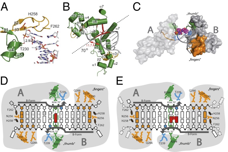
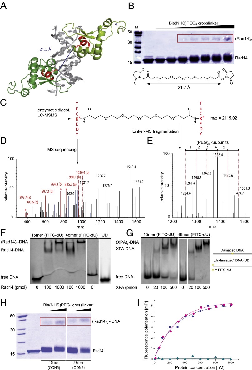
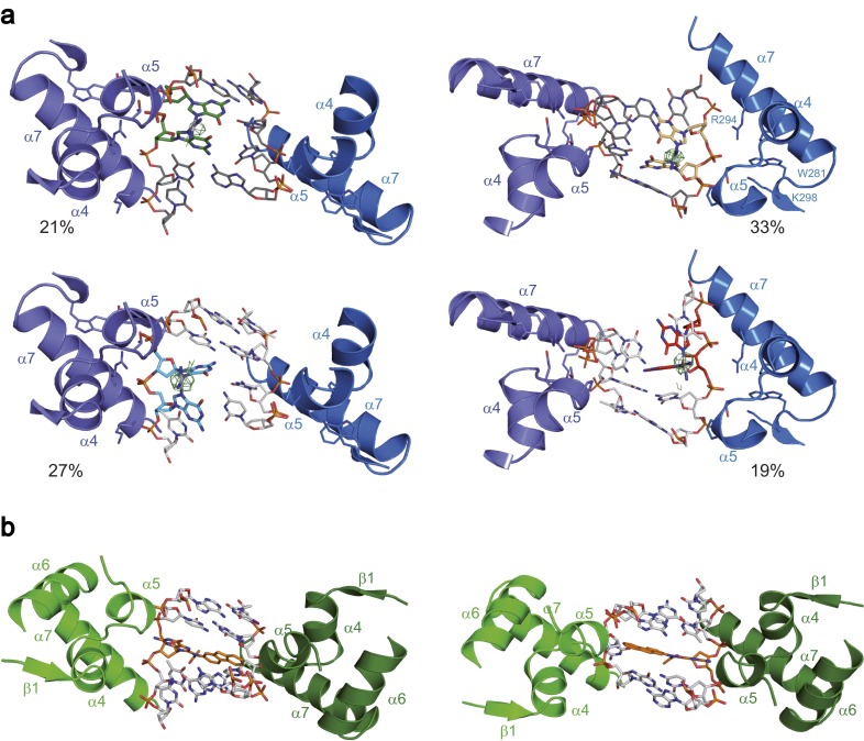
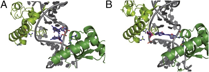
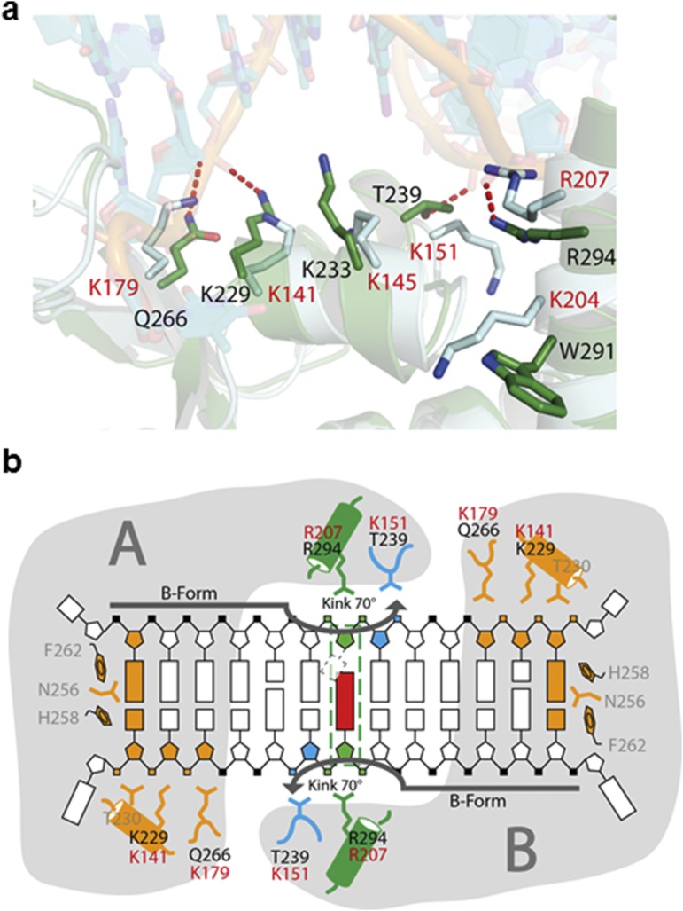
Nucleotide excision repair (NER) is responsible for the removal of a large variety of structurally diverse DNA lesions. Mutations of the involved proteins cause the xeroderma pigmentosum (XP) cancer predisposition syndrome. Although the general mechanism of the NER process is well studied, the function of the XPA protein, which is of central importance for successful NER, has remained enigmatic. It is known, that XPA binds kinked DNA structures and that it interacts also with DNA duplexes containing certain lesions, but the mechanism of interactions is unknown. Here we present two crystal structures of the DNA binding domain (DBD) of the yeast XPA homolog Rad14 bound to DNA with either a cisplatin lesion (1,2-GG) or an acetylaminofluorene adduct (AAF-dG). In the structures, we see that two Rad14 molecules bind to the duplex, which induces DNA melting of the duplex remote from the lesion. Each monomer interrogates the duplex with a β-hairpin, which creates a 13mer duplex recognition motif additionally characterized by a sharp 70° DNA kink at the position of the lesion. Although the 1,2-GG lesion stabilizes the kink due to the covalent fixation of the crosslinked dG bases at a 90° angle, the AAF-dG fully intercalates into the duplex to stabilize the kinked structure.

Keywords: AAF; NER; Rad14; XPA; cisplatin.


Conflict of interest statement

The authors declare no conflict of interest.

Figures

Fig. S1.
Fig. S1.
Binding assays of XPA and Rad14 with different DNA lesions. The indicated amounts of protein were incubated with DNA duplexes (33 fmol) containing the lesions. As control, undamaged dsDNA (UD) was used. Electrophoretic mobility shift assay (EMSA) of XPA with 1,2-GG (A, ODN1 see Table S1), AAF-dG (B, ODN2), and FITC-dU (C, ODN3). EMSA of Rad14 1,2-GG (D, ODN1), AAF-dG (E, ODN2) and UV-lesions [(6, 4)PP and CPD; F, ODN4]. (G) EMSA of Rad14188–302 (Rad14t) with AAF-dG containing DNA (ODN2). (H) Competitive EMSA for the determination of the binding constant of Rad14 to AAF-dG (ODN2). Indicated amounts of Rad14 were incubated with DNA containing AAF-dG (lanes 2–8) and unmodified DNA (lane 9). Competitive DNA was added to the mixture as indicated. The amount of the labeled DNA was counted and assigned as the percentage of bound DNA in the y axis. The x axis shows the logarithmic concentration of the added competition DNA. The curve was fitted as a one-site competition experiment. A binding constant of Kaff = 135 nM (±10 nM SEM) was determined for the affinity of Rad14 to DNA duplexes containing this bulky adduct.
Fig. 1.
Fig. 1.
Overall structures of the Rad14188–302-DNA complexes. (A) Schematic diagrams of the Rad14-AAF-dG and Rad14-1,2-GG complexes showing the different positions where the lesions were observed in orange and red, with the C1’–C1’ distance of the last base pair in the molten part of the structure. For the AAF-dG lesion (Left) two positions of the lesion and for the 1,2-GG lesion (Right) 4 positions of the lesion were observed (Fig. S3). For clarity, only one DNA orientation with the cisplatin lesion is shown here. (B) Schematic representation of the α/β-folding topology of Rad14188–302, with the central 3-stranded β-sheet. (C) Ribbon diagrams of the Rad14-cisplatin lesion DNA complex with the structure of the lesion and the DNA sequence. Rad14 is shown in green and gold (β-hairpin), the DNA backbone is shown in gray, and the cisplatin 1,2-GG lesion is shown in blue. Residues important for DNA kinking (Thr239, His258, Phe262, and Arg294) are shown as sticks. (D) Ribbon diagrams of the Rad14-AAF-dG lesion DNA complex with the structure of the lesion and the DNA sequence. Rad14 is shown in green and gold (β-hairpin), the DNA backbone is shown in gray, and the AAF-dG lesion is shown in blue. Residues important for DNA kinking (Thr239, His258, Phe262 and Arg294) are shown as sticks. In the DNA sequences, the unpaired, partially disordered DNA bases are depicted in light gray. Thymidines replaced by 5-iodo-uracils in the AAF-dG complex are shown in bold.
Fig. S2.
Fig. S2.
Comparison of the human XPA and its yeast homolog Rad14. (A) Structural superposition of the yeast Rad14 DNA-binding domain with the NMR structure (Protein Data Bank ID code 1XPA) of its human homolog (Sequence identity 26.5%, rmsd: 2.2 Å). (B) Sequence alignment of Rad14/XPA homologs. Sequences were aligned with Clustal Omega (76). The secondary structure prediction of the Saccharomyces enzyme was performed with psipred and is shown in the bottom line of the alignment. H represents α-helices, C coils, and E β-strands (77). The boxed area represents the structure shown in the manuscript. The color scheme follows that of Fig. 2. The cysteine residues that build the zinc finger are boxed in yellow.
Fig. 2.
Fig. 2.
Rad14t binds to lesion containing DNA by bending of the DNA. (A) Close up view of the β-hairpin structure with Phe262 and His258 stacking on top of the last base pair. The β-hairpin is shown in orange, Rad14t in green and the DNA as stick model. (B) Bending of the DNA duplex at the lesion position (red) by 70° reduces the P–P distance in the major groove to 11.7 Å. The dotted line represents the pseudo C2 symmetry axis. (C) Schematic representation of the bending process. The β-hairpin is shown in orange, the packing of α7 with Arg294 against the backbone is shown in green, and the stabilization of the bent by Thr239 is shown in blue. (D) Schematic representation of the interaction in the Rad14t-AAF-dG DNA (15mer duplex) complex showing the “fingers” domain mainly formed by the β-hairpin as well as residues Gln266, Lys229, and Thr230 and the “thumb” created by α7 with Arg294. Arg293 (light green) interacts with the backbone only in the AAF-dG structure. The dotted square indicates the dC base opposite the AAF-dG lesion, for which electron density is missing likely due to flexibility. (E) Schematic drawing of the interactions between Rad14t and cisplatin 1,2-GG DNA (16mer duplex) using the same color code as in D.
Fig. 3.
Fig. 3.
Crosslinking experiment of the Rad14t-DNA complex and mass spectrometric analysis of the digested protein dimer. (A) Two Rad14t proteins (light and dark green) bind to one DNA strand containing the AAF-dG lesion (gray). The two lysines at a distance of 21.5 Å in the peptide sequence TECKEDY are highlighted in red. (B) SDS gel of the protein–DNA mixture incubated with increasing amounts of the Bis(NHS)PEG5 crosslinker. (Rad14t)2 bands are boxed in red. The bands were cut out and subjected to enzymatic digestion. (C) Crosslinked peptide sequences after enzymatic digest of the proteins. (D) The MS/MS-spectrum created in an attached HCD-cell of the mass spectrometer reveals the peptide sequence enabling peptide identification (the a- and b-ion series is shown in red, x- and y-ion series is shown in blue). (E) Mass spectrometric analysis revealing the PEG fragments from the crosslinker. The PEG chain fragments with a typical Δm = 44 Da. Electrophoretic mobility shift assay (EMSA) of Rad14fl (F) and XPAfl (G) proteins with a central FITC-dU lesion containing and undamaged DNA (UD; endstanding FITC labeled DNA ODN3 and ODN5, see Table S1). (H) SDS PAGE of the protein–DNA mixture incubated with increasing amounts of the Bis(NHS)PEG5 crosslinker. A 15mer (ODN6) and a 37mer (ODN9) DNA duplex containing an AAF-dG lesion were used. (Rad14t)2 bands are boxed in red. (I) Fluorescence depolarization data showing the DNA binding properties of Rad14t [blue dots: 30mer (ODN10) and purple dots: 15mer (ODN11)] and Rad14tF262A (cyan triangles) to a central FITC-dU lesion containing DNA duplexes.
Fig. S3.
Fig. S3.
Alternative orientations of the DNA bound by two Rad14 proteins. (A) The dinucleotide 1,2-GG lesion occupies four alternative positions in the 13mer duplex wedged between the β-hairpins of the two Rad14 proteins. The occupancy was estimated from the anomalous signal of the platinum atoms. (B) The AAF-dG lesion takes up two opposite positions in the crystal structure.
Fig. 4.
Fig. 4.
Rad14t recognizes lesions that can stabilize kinked structures. (A) Close-up view of the 1,2-GG lesion with the cisplatin unit (blue) intercalated between adjacent base pairs (gray) to stabilize the kinked structure formed by (Rad14t)2 (green). Arg294 is depicted in stick representation with the nitrogens in blue. (B) Close up view of the AAF-dG lesion in complex with Rad14t using the same color code as in A. The flipped out dC was modeled into the structure and is shown in red/blue.
Fig. S4.
Fig. S4.
Van't Hoff plots of Tm−1 vs. ln(Ct/4) derived from the melting curves for the control (undamaged DNA, green) and AAF-dG 15mer duplexes (blue) at concentrations between 2 μM and 7 μM. Ct represents the total oligonucleotide concentration.
Fig. S5.
Fig. S5.
Comparison of Rad14 bound to DNA and human XPA. (A) Superposition of a Rad14 monomer (green) bound to DNA (cyan) with XPA (light blue). Residues that have been implicated in DNA binding for XPA are shown in stick mode. The corresponding residues in Rad14 are also depicted. Human XPA residue numbers are shown in red; Rad14 residue numbers are shown in black. (B) Schematic representation of the Rad14 binding mode to DNA using the scheme introduced in Fig. 2D. Human XPA residues involved in DNA binding are depicted in red.

References

    1. Cleaver JE. Common pathways for ultraviolet skin carcinogenesis in the repair and replication defective groups of xeroderma pigmentosum. J Dermatol Sci. 2000;23(1):1–11. - PubMed
    1. Berneburg M, Lehmann AR. Xeroderma pigmentosum and related disorders: Defects in DNA repair and transcription. Adv Genet. 2001;43:71–102. - PubMed
    1. Naegeli H, Sugasawa K. The xeroderma pigmentosum pathway: Decision tree analysis of DNA quality. DNA Repair (Amst) 2011;10(7):673–683. - PubMed
    1. Sancar A. Mechanisms of DNA excision repair. Science. 1994;266(5193):1954–1956. - PubMed
    1. Geacintov NE, et al. Thermodynamic and structural factors in the removal of bulky DNA adducts by the nucleotide excision repair machinery. Biopolymers. 2002;65(3):202–210. - PubMed

Publication types

MeSH terms