. 2015 Nov 1;407(1):158-72.
doi: 10.1016/j.ydbio.2015.06.015. Epub 2015 Jun 20.

Endocardial Brg1 disruption illustrates the developmental origins of semilunar valve disease

Affiliations

Endocardial Brg1 disruption illustrates the developmental origins of semilunar valve disease

Brynn N Akerberg et al. Dev Biol. .

Abstract

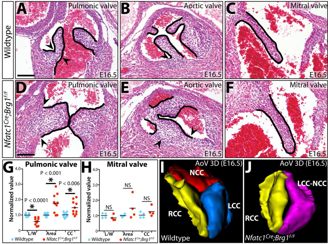
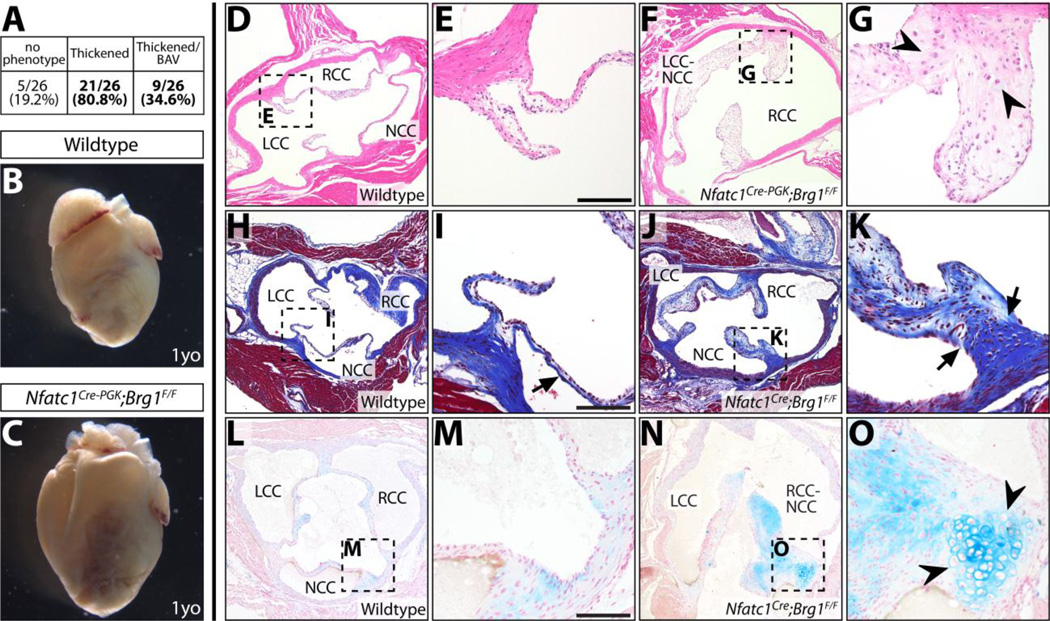
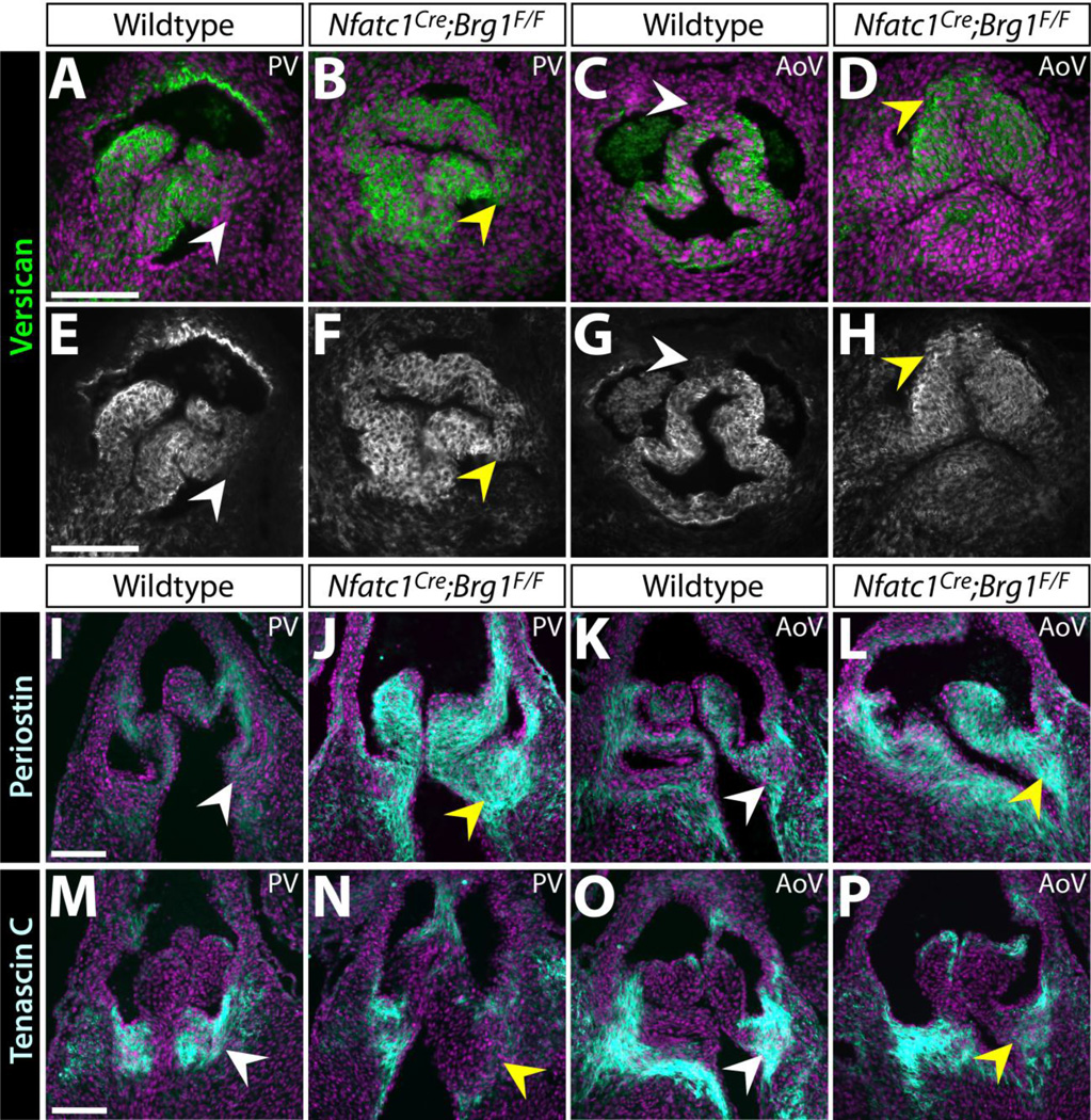
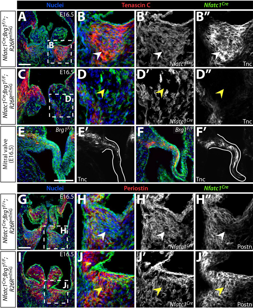
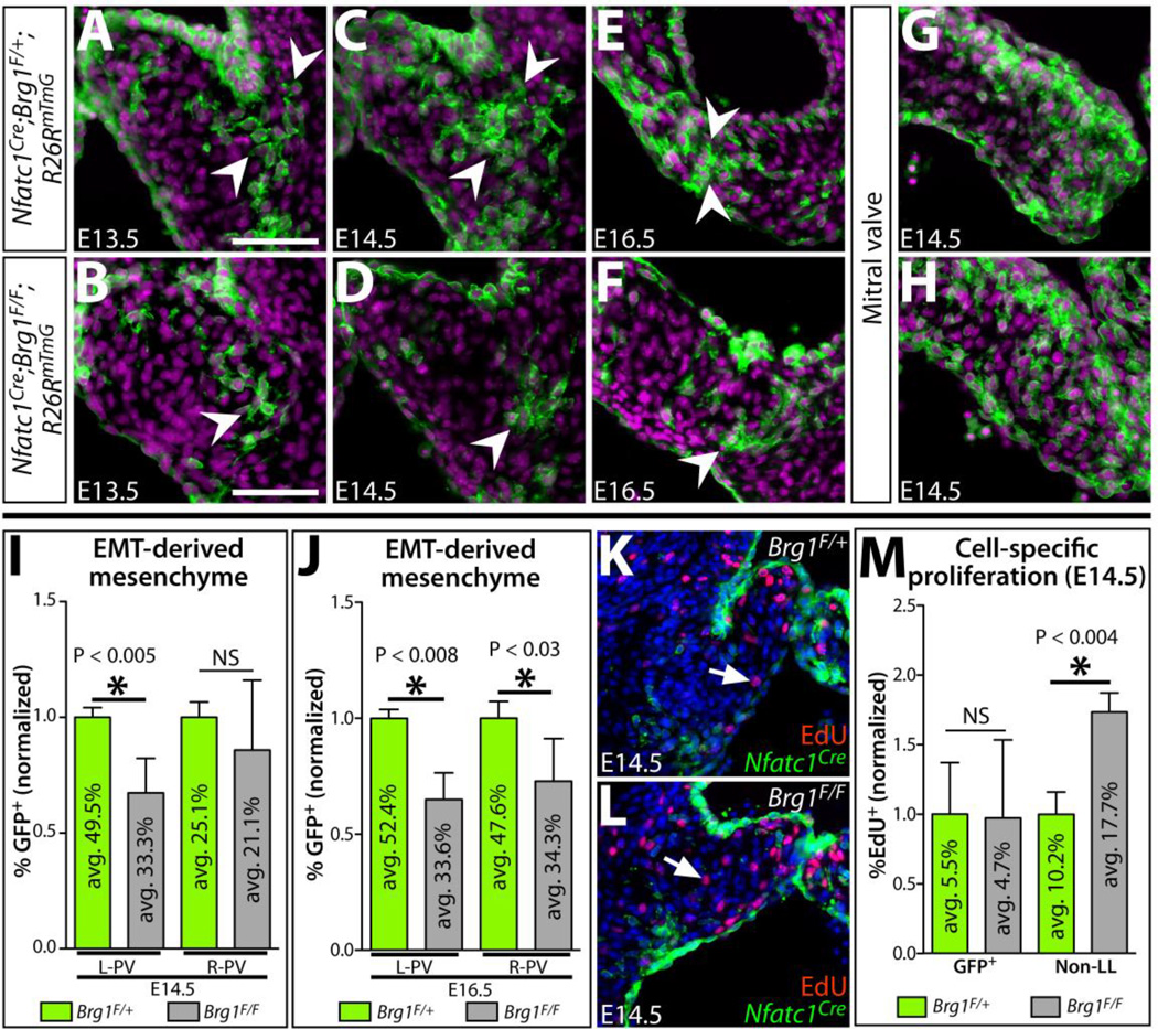
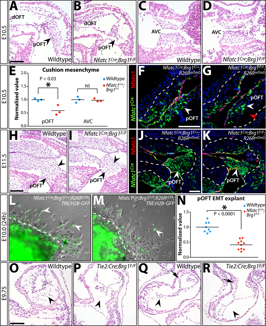
The formation of intricately organized aortic and pulmonic valves from primitive endocardial cushions of the outflow tract is a remarkable accomplishment of embryonic development. While not always initially pathologic, developmental semilunar valve (SLV) defects, including bicuspid aortic valve, frequently progress to a disease state in adults requiring valve replacement surgery. Disrupted embryonic growth, differentiation, and patterning events that "trigger" SLV disease are coordinated by gene expression changes in endocardial, myocardial, and cushion mesenchymal cells. We explored roles of chromatin regulation in valve gene regulatory networks by conditional inactivation of the Brg1-associated factor (BAF) chromatin remodeling complex in the endocardial lineage. Endocardial Brg1-deficient mouse embryos develop thickened and disorganized SLV cusps that frequently become bicuspid and myxomatous, including in surviving adults. These SLV disease-like phenotypes originate from deficient endocardial-to-mesenchymal transformation (EMT) in the proximal outflow tract (pOFT) cushions. The missing cells are replaced by compensating neural crest or other non-EMT-derived mesenchyme. However, these cells are incompetent to fully pattern the valve interstitium into distinct regions with specialized extracellular matrices. Transcriptomics reveal genes that may promote growth and patterning of SLVs and/or serve as disease-state biomarkers. Mechanistic studies of SLV disease genes should distinguish between disease origins and progression; the latter may reflect secondary responses to a disrupted developmental system.

Keywords: BAF complex; Bicuspid aortic valve; Brg1; Chromatin remodeling; Endocardial cushions; Endocardial-to-mesenchymal transformation; Outflow tract; Semilunar valves; Valve disease.


Figures

Figure 1
Figure 1. Endocardial-lineage Brg1 deficient embryos exhibit semilunar valve defects
(A–F) Hematoxylin and eosin (H&E) stained sections from E16.5 wildtype and Nfatc1Cre;Brg1F/Fembryos showing indicated valves. Arrowheads mark the basal cusp regions of the semilunar valves. (G, H) Morphometric analyses of pulmonic and mitral valves from E16.5 wildtype and Nfatc1Cre;Brg1F/F embryos. Each data point represents one valve, with values normalized to the mean of the wildtype samples from individual litters. Asterisks indicate a significant difference (P < 0.05, Student’s two-tailed t-tests). (I, J) 3-dimensional rendering of serial sections of aortic valves from E16.5 wildtype and Nfatc1Cre;Brg1F/F littermate embryos. AoV: aortic valve; L/W: length-to-width; CC: mesenchymal cell count; NS, not statistically significant; LCC (blue): left coronary cusp; NCC (red): non-coronary cusp; RCC (yellow): right coronary cusp; LCC-NCC (purple): bicuspid fusion between left and non-coronary cusps. Scale bars: 100 µm.
Figure 2
Figure 2. Endocardial-lineage Brg1 deficient adult mice develop aortic valve disease
(A) Fraction of adult Nfatc1Cre-PGK;Brg1F/F or Nfatc1Cre;Brg1F/F mice with 1) thickened/myxomatous and/or 2) bicuspid aortic valve. 0/26 littermate control animals had abnormal aortic valves. (B, C) Whole mount anterior view of hearts from one year old (1yo) wildtype and Nfatc1Cre-PGK;Brg1F/F littermate mice. (D–G) H&E stained wildtype (D, E) and Nfatc1Cre-PGK;Brg1F/F (F, G) aortic valve sections. (H–K) Adult aortic valve sections stained using Masson’s Trichrome stain (collagen deposition in blue). Wildtype (H, I) and Nfatc1Cre;Brg1F/F (J, K) samples are shown. Black arrows mark mislocalized collagen. (L–O) Von Kossa and Alcian Blue (counterstained with Nuclear Fast Red) stained tissue sections from wildtype (L, M) and Nfatc1Cre;Brg1F/F (N, O) adult aortic valves. Black arrowheads indicate cartilaginous nodules. LCC: left coronary cusp; NCC: non-coronary cusp; RCC: right coronary cusp; LCC/RCC-NCC: bicuspid fusion between left/right and non-coronary cusps. Scale bars: 100 µm.
Figure 3
Figure 3. Altered localization and levels of extracellular matrix proteins in Nfatc1Cre;Brg1F/F semilunar valves
(A–H) Widefield fluorescence images of anti-Versican (Vcan) antibody stained sections of E16.5 wildtype and Nfatc1Cre;Brg1F/F embryos. (I–P) Representative confocal imaged cryosections of E16.5 wildtype and Nfatc1Cre;Brg1F/F semilunar valves fluorescently immunostained for Periostin (Postn) (I–L) or Tenascin C (Tnc) (M–P) (n=3, paired control and Brg1-deficient valves). Pulmonic (PV) or aortic (AoV) valves are shown as indicated. ECM proteins are shown in green/gray scale (A–D) or cyan (I–P). Hoechst-stained nuclei are blue. White arrowheads mark areas of normal protein expression. Yellow arrowheads show regions with broadened (Vcan), broadened/increased (Postn) or decreased (Tnc) levels. Scale bars: 100 µm.
Figure 4
Figure 4. Disorganized extracellular matrix in endocardial Brg1 deficient semilunar valves reflects an altered organization of distinct mesenchymal lineages
Immunofluorescence images of E16.5 control (Nfatc1Cre;Brg1F/+;R26RmTmG) or endocardial Brg1-deficient (Nfatc1Cre;Brg1F/F;R26RmTmG) embryo sections simultaneously stained for GFP to monitor Nfatc1Cre-lineage cells and either Tenascin C (Tnc) (A–F) or Periostin (Postn) (G–J). Pulmonic valves (A–D, G–J) and mitral valves (E–F) are shown. All panels are confocal images except (E–F), which are widefield images. Overlay images show Tnc or Postn in red, GFP indicating Nfatc1Cre-lineage cells in green, and Hoechst-stained nuclei in blue. Magnified regions are outlined in dashed boxes. White arrowheads denote normal ECM localization and yellow arrowheads represent either deficient (Tnc) or ectopic (Postn) ECM protein. Scale bars: 100 µm.
Figure 5
Figure 5. A depleted pool of EMT-derived mesenchyme in endocardial Brg1-deficient semilunar valves is replaced by increased proliferation of other mesenchymal lineage(s)
(A–H) Sections of Nfatc1Cre;Brg1F/+;R26RmTmG and Nfatc1Cre;Brg1F/F;R26RmTmG embryos (pulmonic valve except mitral valve in (G, H); genotype and age as indicated) immunostained for GFP (Nfatc1Cre-lineage derived cells, green) and nuclei (Hoechst-stained, purple). Arrowheads indicate EMT-derived mesenchymal cells at the pulmonic valve cusp base near the cushion-myocardium boundary. (I, J) Quantification of the relative contribution of Nfatc1Cre-lineage pulmonic valve mesenchyme in control (Brg1F/+) and endocardial Brg1-deficient embryos (Brg1F/F) at E14.5 (I) and E16.5 (J). Left (L-PV) and right (R-PV) cusps are scored separately. The percentile fraction of Nfatc1Cre-lineage mesenchyme for each embryo is normalized to the mean of corresponding littermate wildtype samples. The mean absolute percentage of labeled and unlabeled cells is shown within individual bars of the chart (n=5). (K–L) E14.5 wildtype (Nfatc1Cre;Brg1F/+;R26RmTmG) (K) and Nfatc1Cre;Brg1F/F;R26RmTmG (L) pulmonic valve sections stained with GFP (Nfatc1Cre-lineage, green), EdU-incorporating proliferating cells (red), and Hoechst (nuclei, blue). White arrows denote EdU+, non-Nfatc1Cre-lineage mesenchymal cells. (M) Quantitation of the proliferation rate of Nfatc1Cre- and other-lineage pulmonic valve mesenchyme in E14.5 control and Nfatc1Cre;Brg1F/F embryos. The mean absolute fraction of EdU-incorporated cells for each condition is shown. For all graphs, error bars show one standard deviation of the mean. Asterisks indicate a significant difference (P < 0.05, two-tailed Student’s t-tests). NS: not significant; GFP+Nfatc1Cre-lineage labeled mesenchyme; Non-LL: non-Nfatc1Cre-lineage labeled mesenchyme. Scale bars: 100 µm.
Figure 6
Figure 6. Reduced EMT-derived semilunar valve mesenchyme i7n Nfatc1Cre;Brg1F/F embryos originates from deficient pOFT EMT, a process that generally requires Brg1 function
(A–D) H&E stained sagittal sections of E10.5 wildtype and Nfatc1Cre;Brg1F/F embryos. The proximal (pOFT) and distal (dOFT) outflow tract (A, B) and atrioventricular canal (AVC) cushions (C, D) are shown. Arrowheads indicate pOFT mesenchyme. (E) Scoring of mesenchymal cells populating the pOFT or AVC cushions in E10.5 wildtype and Nfatc1Cre;Brg1F/F embryos. Each point shows one embryo and all values are normalized to the mean of littermate wildtype samples. Asterisk denotes a significant difference (two-tailed Student’s t-tests). (F-G, J-K) Widefield fluorescence images of OFT regions from transverse sectioned E10.5 (F, G) or sagittal sectioned E11.5 (J, K) embryos. Tissue is immunostained for GFP (for Nfatc1Cre-lineage tracing, green) and Nfatc1 (endocardium, red). Nuclei are stained with Hoechst (blue). Control (Nfatc1Cre;Brg1F/+;R26RmTmG; F, J) and Nfatc1Cre;Brg1F/F;R26RmTmG (G, K) embryos are shown. (H, I) H&E stained sagittal sections of E11.5 wildtype and Nfatc1Cre;Brg1F/F embryos. Arrowheads indicate pOFT mesenchyme. (L, M) Brightfield and fluorescence overlaid images of collagen gel OFT explants from E10.0 control and Nfatc1Cre;Brg1F/F embryos (24 hours post-dissection). All embryos additionally carry R26RrtTA and TRE:H2B–GFP transgenes and explants are treated with doxycycline to induce H2B–GFP expression (to label Nfatc1Cre-lineage mesenchymal cells, green). White arrowheads denote EMT-derived (GFP+) mesenchymal cells. (N) Quantification of Nfatc1Cre-lineage mesenchymal cells migrating through the collagen matrix. Each point represents one explanted OFT. Values are normalized to the mean of littermate wildtype explants. The asterisk indicates statistical significance (two-tailed Student’s t-test). (O–R) Endocardial Brg1 is required for all cushion EMT. H&E stained sections showing AVC (O,P) and pOFT (Q,R) cushions of wildtype and Tie2:Cre;Brg1F/F E9.75 littermate embryos. Arrowheads denote either the presence or a depleted/absent pool of endocardial-derived cushion mesenchymal cells. Arrows in (Q, R) show neural crest-origin mesenchyme within the dOFT cushions. Scale bars: 100 µm.
Figure 7
Figure 7. RNA-seq identifies novel semilunar valve expressed transcripts misexpressed in endocardial lineage Brg1 deficient valves
(A) Select differentially expressed genes (DEGs) identified from an RNA-seq analysis comparing two paired samples of wildtype and Nfatc1Cre;Brg1F/F dissected E14.5 heart valve tissue. 26 DEGs (green-upregulated; red-downregulated) are listed with fold change and false discovery rate (FDR). Transcripts of particular interest are highlighted. (B,C) Quantitative RT-PCR expression studies of the highlighted DEGs in (A) E14.5 (B) and E16.5 (C) valves. Bar heights represent the log-base-2 fold change in gene expression (linear mean fold change value indicated below bars) between Nfatc1Cre;Brg1F/F and wildtype tissue. Error bars represent one standard deviation with the additional error bars centered along the x-axis showing the variation between wildtype samples. Double asterisks indicate a significant difference including a Bonferroni correction (one-tailed Student’s t-test, n=7, P < 0.007); single asterisk indicates a P < 0.05. (D–K) RNA in situ hybridizations for indicated transcripts on pulmonic valve sections of E13.5 wildtype or littermate Nfatc1Cre;Brg1F/F embryos. Arrowheads highlight areas of highest transcript expression. Scale bar: 100 µm.

References

    1. Angelini A, Ho SY, Anderson RH, Devine WA, Zuberbuhler JR, Becker AE, Davies MJ. The morphology of the normal aortic valve as compared with the aortic valve having two leaflets. J Thorac Cardiovasc Surg. 1989;98:362–367. - PubMed
    1. Boles NC, Hirsch SE, Le S, Corneo B, Najm F, Minotti AP, Wang Q, Lotz S, Tesar PJ, Fasano CA. NPTX1 Regulates Neural Lineage Specification from Human Pluripotent Stem Cells. Cell Reports. 2014;6:724–736. - PubMed
    1. Bosse K, Hans CP, Zhao N, Koenig SN, Huang N, Guggilam A, LaHaye S, Tao G, Lucchesi PA, Lincoln J, et al. Endothelial nitric oxide signaling regulates Notch1 in aortic valve disease. Journal of Molecular and Cellular Cardiology. 2013;60:27–35. - PMC - PubMed
    1. Broom ND. The observation of collagen and elastin structures in wet whole mounts of pulmonary and aortic leaflets. J Thorac Cardiovasc Surg. 1978;75:121–130. - PubMed
    1. Bultman S, Gebuhr T, Yee D, La Mantia C, Nicholson J, Gilliam A, Randazzo F, Metzger D, Chambon P, Crabtree G, et al. A Brg1 null mutation in the mouse reveals functional differences among mammalian SWI/SNF complexes. Molecular Cell. 2000;6:1287–1295. - PubMed

Publication types

LinkOut - more resources