Review
. 2015 Jun 22;8(1):a020479.
doi: 10.1101/cshperspect.a020479.

Oligodendrocytes: Myelination and Axonal Support

Affiliations
Review

Oligodendrocytes: Myelination and Axonal Support

Mikael Simons et al. Cold Spring Harb Perspect Biol. .

Abstract

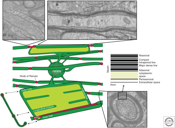
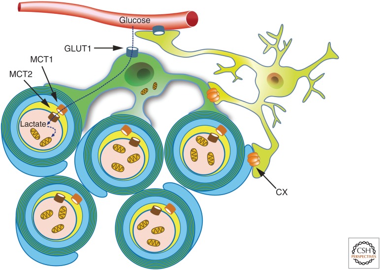
Myelinated nerve fibers have evolved to enable fast and efficient transduction of electrical signals in the nervous system. To act as an electric insulator, the myelin sheath is formed as a multilamellar membrane structure by the spiral wrapping and subsequent compaction of the oligodendroglial plasma membrane around central nervous system (CNS) axons. Current evidence indicates that the myelin sheath is more than an inert insulating membrane structure. Oligodendrocytes are metabolically active and functionally connected to the subjacent axon via cytoplasmic-rich myelinic channels for movement of macromolecules to and from the internodal periaxonal space under the myelin sheath. This review summarizes our current understanding of how myelin is generated and also the role of oligodendrocytes in supporting the long-term integrity of myelinated axons.


Figures

Figure 1.
Figure 1.
Morphology of oligodendroglia in the cerebellum of a cat. (Top right) Cellular processes and branches follow the orientation of the nerve fibers and form complex wraps as shown in del Río-Hortega (1921). (Top left) White matter of a newborn human brain: A, Elongated interfascicular cells filled with spherical granules of variable size; B and C, round granular cells; D, astrocytes with long processes; and E, nucleus of a microglia as shown in del Río-Hortega (1921). (Bottom left) The four types of oligodendrocytes recognized by del Río-Hortega. (Bottom right) Oligodendrocytes expressing proteolipid protein (PLP)–enhanced green fluorescent protein (EGFP) in transgenic mice. Because of its bulky EGFP tag, most of it is found within the cytoplasmic-rich spaces or myelin, including the myelinic channels.
Figure 2.
Figure 2.
Schematic view of myelinating oligodendrocytes in its wrapped and conceptually “unrolled” state. Electron micrographs show (left) paranodal loops, a longitudinal view (right), and a cross-section view (bottom) of myelin. (From Aggarwal et al. 2011a; modified, with permission, from Elsevier Limited © 2011.)
Figure 3.
Figure 3.
Model showing the direction of how myelin wraps around the axon. There are two motions: the wrapping of the leading edge at the inner tongue around the axon underneath the previously deposited membrane and the lateral extension of myelin membrane layers toward the nodal regions (for details, see Snaidero et al. 2014).
Figure 4.
Figure 4.
Schematic depiction of an oligodendrocyte that takes up blood-derived glucose and delivers glycolysis products (pyruvate/lactate) via monocarboxylate transporters (MCT1 and MCT2) to myelinated axons. Oligodendrocytes and myelin membranes are also coupled by gap junctions to astrocytes and, thus, indirectly to the blood–brain barrier. CX, Connexin. (From Saab et al. 2013; modified, with permission, from Elsevier Limited © 2013.)

References

    1. Abrams CK, Scherer SS. 2012. Gap junctions in inherited human disorders of the central nervous system. Biochim Biophys Acta 1818: 2030–2047. - PMC - PubMed
    1. Aggarwal S, Yurlova L, Simons M. 2011a. Central nervous system myelin: Structure, synthesis and assembly. Trends Cell Biol 21: 585–593. - PubMed
    1. Aggarwal S, Yurlova L, Snaidero N, Reetz C, Frey S, Zimmermann J, Pahler G, Janshoff A, Friedrichs J, Muller DJ, et al. 2011b. A size barrier limits protein diffusion at the cell surface to generate lipid-rich myelin-membrane sheets. Dev Cell 21: 445–456. - PubMed
    1. Aggarwal S, Snaidero N, Pahler G, Frey S, Sanchez P, Zweckstetter M, Janshoff A, Schneider A, Weil MT, Schaap IA, et al. 2013. Myelin membrane assembly is driven by a phase transition of myelin basic proteins into a cohesive protein meshwork. PLoS Biol 11: e1001577. - PMC - PubMed
    1. Ainger K, Avossa D, Morgan F, Hill SJ, Barry C, Barbarese E, Carson JH. 1993. Transport and localization of exogenous myelin basic protein mRNA microinjected into oligodendrocytes. J Cell Biol 123: 431–441. - PMC - PubMed

Publication types