. 2015 Aug;36(2):345-54.
doi: 10.3892/ijmm.2015.2258. Epub 2015 Jun 23.

Puerarin attenuates glucocorticoid-induced apoptosis of hFOB1.19 cells through the JNK- and Akt-mediated mitochondrial apoptotic pathways

Affiliations

Puerarin attenuates glucocorticoid-induced apoptosis of hFOB1.19 cells through the JNK- and Akt-mediated mitochondrial apoptotic pathways

Dongdong Yu et al. Int J Mol Med. 2015 Aug.

Abstract

Puerarin is an active component of Pueraria lobata, which is a commonly used Chinese herbal medicine for the treatment of osteoporosis. The present study aimed to evaluate the osteoprotective effect of puerarin on glucocorticoid (GC)-induced apoptosis of osteoblasts in vitro. The effects of puerarin on dexamethasone (DEX)-induced cell apoptosis were assessed using enzyme-linked immunosorbent assay and a terminal deoxynucleotidyl transferase dUTP nick-end labeling assay, and found that the viability of hFOB1.19 cells was significantly increased following exposure to between 10(-6) and 10(-10) M puerarin, with a maximal anti-apoptotic effect at a concentration of 10(-8) M. In addition, compared with the control group, puerarin upregulated the transcription and protein levels of B-cell lymphoma-2 and downregulated B-cell-associated X protein in the hFOB1.19 cells. Puerarin attenuated the DEX-induced release of cytochrome c and cleavage of caspase-3, and treatment with puerarin inhibited the c-Jun N-terminal kinase (JNK) pathway and activated the phosphoinositide 3-kinase (PI3K)/Akt pathway in the hFOB1.19 cells. Furthermore, the Akt inhibitor, LY294002, partly eliminated the protective effect of puerarin on DEX-induced apoptosis, and puerarin combined with the JNK inhibitor, SP600125, suppressed DEX-induced apoptosis to a lesser extent than in the cells treated with SP600125 alone. These results suggested that the JNK and PI3K/Akt signaling pathways mediate the inhibitory effects of puerarin on apoptosis in the hFOB1.19 cells. In conclusion, puerarin prevented DEX-induced apoptosis of hFOB1.19 cells via inhibition of the JNK pathway and activation of the PI3K/Akt signaling pathway in the cells, dependent on the mitochondrial apoptotic pathway. These results support puerarin as a promising target in the treatment of GC-induced osteoporosis.


Figures

Figure 1
Figure 1
Puerarin promotes hFOB1.19 cell proliferation. Cells proliferation was measured using an MTS detection kit. The cells were treated with 0 M and 10−6–10−10 M puerarin for 3, 12 and 24 h. Treatment with 10−8 and 10−9 M puerarin significantly increased cell proliferation. Data are presented as the mean ± standard deviation. *P<0.05, vs control. OD, optical density.
Figure 2
Figure 2
DEX inhibits hFOB1.19 cell proliferation. Cell proliferation was measured using an MTS detection kit following pretreatment of the cells with 0 M and 10−5–10−9 M DEX for 24, 48 and 72 h. Treatment with 10−5 and 10−6 M DEX significantly reduced cell proliferation. Data are presented as the mean ± standard deviation. *P<0.05, vs. control. DEX, dexamethasone; OD, optical density.
Figure 3
Figure 3
Purearin alleviates DEX-induced hFOB1.19 cell apoptosis. Cell apoptosis was measured using an enzyme-linked immunosorbent assay kit following pretreatment of the cells with 0 M and 10−6–10−10 M puerarin for 3 h. The cells were then incubated with DEX (10−5 M) for 48 h. Treatment with 10−8 and 10−9 M puerarin significantly reduced cell apoptosis. Data are presented as the mean ± standard deviation. *P<0.05, vs. DEX. DEX, dexamethasone.
Figure 4
Figure 4
Effects of puerarin on apoptosis in hFOB1.19 cells, determined using a TUNEL assay. (A) Cells were pretreated with puerarin (0 or 10−8–10−9 M) for 3 h, and were then incubated with DEX (10−5 M) for 48 h. The number of apoptotic cells were determined using TUNEL staining and observed under a fluorescence microscope; original magnification, ×100. (B) Percentages of positive cells were calculated in six randomly selected fields as the apoptotic index using the following equation: AI = (number of positive cells/total number of cells) × 100%. Arrows indicate apoptotic cells. Data are presented as the mean ± standard deviation (n=3). *P<0.05, vs. DEX; #P<0.05, vs. control. DEX, dexamethasone; TUNEL, terminal deoxynucleotidyltransferase-mediated dUTP nick-end labeling; DAPI, 4-Diamino-2-phenylindole.
Figure 5
Figure 5
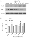
Effects of puerarin on DEX-induced changes in of Bcl-2 and Bax. (A) Cells were treated with puerarin (0, 10−7, 10−8 and 10−9 M) for 3 h prior to incubation with DEX (10−5 M) for 48 h. Western blot analyses for Bcl-2, Bax and β-actin was performed. (B) Relative expression levels of Bcl-2 and Bax compared with β-actin. Data are presented as the mean ± standard deviation (n=3). *P<0.05, vs. DEX; #P<0.05, vs. control. DEX, dexamethasone; Bcl-2, B-cell lymphoma-2; Bax, B-cell-associated X protein.
Figure 6
Figure 6
Effects of puerarin on the expression levels of caspase-3 and cytochrome c in the hFOB1.19 cells. (A) Cells were treated with puerarin (0, 10−7, 10−8 and 10−9 M) for 3 h prior to incubation with DEX (10−5 M) for 48 h. Western blot analyses were performed to detect the expression levels of caspase-3, cleaved caspase-3, cytochrome c, and β-actin. (B) Relative expression levels of cleaved caspase-3 and cytochrome c were compared with the expression levels of total caspase-3 and β-actin. Data are presented as the mean ± standard deviation (n=3). *P<0.05, vs. DEX; #P<0.05, vs. control. DEX, dexamethasone.
Figure 7
Figure 7
RT-qPCR analysis of caspase-3, Bax and Bcl-2. The cells were treated with puerarin (0, 10–7, 10−8 and 10−9 M) for 3 h prior to incubation with DEX (10−5 M) for 48 h. RT-qPCR was performed to analyzed expression levels of caspase-3, Bax and Bcl-2. Data are presented as the mean ± standard deviation. *P<0.05, vs. DEX; #P<0.05, vs. control. RT-qPCR, reverse transcription-quantitative polymerase chain reaction; DEX, dexamethasone; Bcl-2, B-cell lymphoma-2; Bax, B-cell-associated X protein.
Figure 8
Figure 8
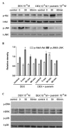
Effects of puerarin on the phosphorylation of JNK and Akt in hFOB1.19 cells. (A and C) Cells were treated with DEX (10−5 M) alone or in combination with puerarin (10−8 M) for 5, 30 and 60 min. Western blot analysis was performed to detect the expression levels of p-JNK, JNK, p-Akt, Akt, p-ERK and ERK. (B) Relative expression levels of p-Akt and p-JNK compared with t-Akt and t-JNK were analyzed, respectively. Data are presented as the mean ± standard deviation (n=3). *P<0.05, vs. DEX; #P<0.05, vs. control. JNK, c-Jun N-terminal kinase; ERK, extracellular signal-regulated kinase; p-, phosphorylated; t-, total.
Figure 9
Figure 9
Effects of LY294002 and SP600125 on the expression levels of p-Akt and p-JNK in hFOB1.19 cells. (A) Cells were treated with DEX (10−5 M), DEX (10−5 M) + LY294002 (10−5 M), or DEX (10−5 M) + puerarin (10−8 M) + LY294002 (10−5 M). Expression levels of p-Akt and t-Akt were detected using western blot analysis. (B) Cells were treated with DEX (10−5 M), DEX (10−5 M) + SP600125 (10−5 M), or DEX (10−5 M) + puerarin (10−8 M) + SP600125 (10−5 M). Expression levels of p-JNK and t-JNK were detected using western blot analysis. Data are presented as the mean ± standard deviation. *P<0.05, vs. DEX. JNK, c-Jun N-terminal kinase; p-, phosphorylated; t-, total.
Figure 10
Figure 10
Effects of JNK and Akt on the apoptosis of hFOB1.19 cells. (A) Cells were treated with puerarin (10−8 M), SP600125 (10−5 M) or LY294002 (10−5 M) for 3 h prior to incubation with DEX (10−5 M) for 48 h. Flow cytometric analysis was performed following Annexin V-FITC/PI double staining. The upper left quadrant represents dead cells or debris, the upper right quadrant represents the late stage of apoptotic cells, the lower right quadrant represents the early stage of apoptotic cells and the lower left quadrant represented normal cells. (B) Levels of apoptosis of the hFOB1.19 cells were analyzed. Data are presented as the mean ± standard deviation. *P<0.01, vs. DEX; #P<0.05, vs. control. DEX, dexamethasone; FITC, fluorescein isothiocyanate; PI, propidium iodide.

References

    1. Canalis E. Clinical review 83: Mechanisms of glucocorticoid action in bone: implications to glucocorticoid-induced osteoporosis. J Clin Endocrinol Metab. 1996;81:3441–3447. - PubMed
    1. Manelli F, Giustina A. Glucocorticoid-induced osteoporosis. Trends Endocrinol Metab. 2000;11:79–85. doi: 10.1016/S1043-2760(00)00234-4. - DOI - PubMed
    1. Tamura Y, Okinaga H, Takami H. Glucocorticoid-induced osteoporosis. Biomed Pharmacother. 2004;58:500–504. doi: 10.1016/j.biopha.2004.08.018. - DOI - PubMed
    1. Weinstein RS, Jilka RL, Parfitt AM, Manolagas SC. Inhibition of osteoblastogenesis and promotion of apoptosis of osteoblasts and osteocytes by glucocorticoids. Potential mechanisms of their deleterious effects on bone. J Clin Invest. 1998;102:274–282. doi: 10.1172/JCI2799. - DOI - PMC - PubMed
    1. Wu L, Qiao H, Li Y, Li L. Protective roles of puerarin and Danshensu on acute ischemic myocardial injury in rats. Phytomedicine. 2007;14:652–658. doi: 10.1016/j.phymed.2007.07.060. - DOI - PubMed

Publication types

MeSH terms