Review
. 2015 Jun 22;5(8):a022863.
doi: 10.1101/cshperspect.a022863.

Glutamatergic Mechanisms Associated with Seizures and Epilepsy

Affiliations
Review

Glutamatergic Mechanisms Associated with Seizures and Epilepsy

Melissa Barker-Haliski et al. Cold Spring Harb Perspect Med. .

Abstract

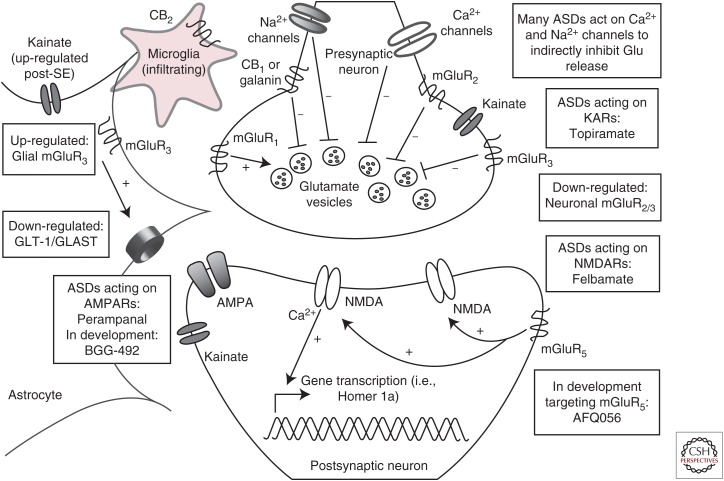
Epilepsy is broadly characterized by aberrant neuronal excitability. Glutamate is the predominant excitatory neurotransmitter in the adult mammalian brain; thus, much of past epilepsy research has attempted to understand the role of glutamate in seizures and epilepsy. Seizures induce elevations in extracellular glutamate, which then contribute to excitotoxic damage. Chronic seizures can alter neuronal and glial expression of glutamate receptors and uptake transporters, further contributing to epileptogenesis. Evidence points to a shared glutamate pathology for epilepsy and other central nervous system (CNS) disorders, including depression, which is often a comorbidity of epilepsy. Therapies that target glutamatergic neurotransmission are available, but many have met with difficulty because of untoward adverse effects. Better understanding of this system has generated novel therapeutic targets that directly and indirectly modulate glutamatergic signaling. Thus, future efforts to manage the epileptic patient with glutamatergic-centric treatments now hold greater potential.


Figures

Figure 1.
Figure 1.
The glutamatergic tripartite synapse. Excitatory afferents project from cortical or hippocampal regions, releasing glutamate into the synaptic cleft. Under normal conditions, synaptic glutamate can signal through ionotropic α-amino-3-hydroxy-5-methyl-4-isoxazolepropionic acid receptors (AMPARs), N-methyl-d-aspartate receptors (NMDARs), and kainate receptors (KARs), or through metabotropic mGluR I, II, or III class receptors. Synaptic glutamate is subject to reuptake by astrocytes via the glutamate transporters, glutamate transporter 1 (GLT)-1 and l-glutamate/l-aspartate transporter (GLAST). In epilepsy, GLT-1 and GLAST expression is down-regulated, an effect that can contribute to further excitotoxic damage under conditions of chronic seizure-induced glutamate release. Currently approved antiseizure drugs (ASDs) target some aspects of the glutamatergic synapse and more compounds are currently in development to selectively modulate glutamatergic signaling through mGluRs, as well as glial receptors and transporters. Astrocyte-specific strategies to augment GLT-1/GLAST expression in epilepsy, and thereby a decrease in extraneous synaptic levels of excitotoxic glutamate, is also an area of active investigation for therapeutic intervention. Additional areas of therapeutic development include mGluR targeting agents, cannibinoid (CB) receptor targeting compounds, and neuropeptide receptors, all of which are hypothesized to directly and indirectly modulate glutamatergic signaling. SE, Status epilepticus; mGluR, metabotropic glutamate receptor.
Figure 2.
Figure 2.
Representative photomicrographs from in situ hybridization histochemistry. Homer 1a messenger RNA (mRNA) expression in dorsal hippocampus of male Sprague–Dawley rats (A) 60 min, and (B) 120 min following maximal electroshock (MES) stimulation (White et al. 1995b; Loscher 1997). Homer 1a mRNA shows robust activity-dependent expression 60 min and 120 min following MES (Brakeman et al. 1997) in dorsal hippocampus. In situ hybridization histochemical labeling for Homer 1a mRNA was conducted as previously described for Arc mRNA (Barker-Haliski et al. 2012), with complementary DNA (cDNA) plasmids for Homer 1a provided by Dr. Kristen Keefe, University of Utah. Images were acquired at 2.5× magnification using a Zeiss Axio Imager.A1 microscope and Axiovision V.4.5 imaging software. Green is Homer 1a mRNA and blue is DAPI nuclear counterstain (Life Technologies, Norwalk, CT).

References

    1. Abulseoud OA, Miller JD, Wu J, Choi DS, Holschneider DP. 2012. Ceftriaxone upregulates the glutamate transporter in medial prefrontal cortex and blocks reinstatement of methamphetamine seeking in a condition place preference paradigm. Brain Res 1456: 14–21. - PMC - PubMed
    1. Alexander GM, Godwin DW. 2006. Unique presynaptic and postsynaptic roles of group II metabotropic glutamate receptors in the modulation of thalamic network activity. Neuroscience 141: 501–513. - PubMed
    1. Alexopoulos H, Kosmidis ML, Dalmau J, Dalakas MC. 2011. Paraneoplastic anti-NMDAR encephalitis: Long-term follow-up reveals persistent serum antibodies. J Neurol 258: 1568–1570. - PubMed
    1. Altar CA, Laeng P, Jurata LW, Brockman JA, Lemire A, Bullard J, Bukhman YV, Young TA, Charles V, Palfreyman MG. 2004. Electroconvulsive seizures regulate gene expression of distinct neurotrophic signaling pathways. J Neurosci 24: 2667–2677. - PMC - PubMed
    1. Anggono V, Huganir RL. 2012. Regulation of AMPA receptor trafficking and synaptic plasticity. Curr Opin Neurobiol 22: 461–469. - PMC - PubMed