. 2015 Jul 27;43(13):6486-99.
doi: 10.1093/nar/gkv633. Epub 2015 Jun 22.

SAMHD1 is a single-stranded nucleic acid binding protein with no active site-associated nuclease activity

Affiliations

SAMHD1 is a single-stranded nucleic acid binding protein with no active site-associated nuclease activity

Kyle J Seamon et al. Nucleic Acids Res. .

Abstract

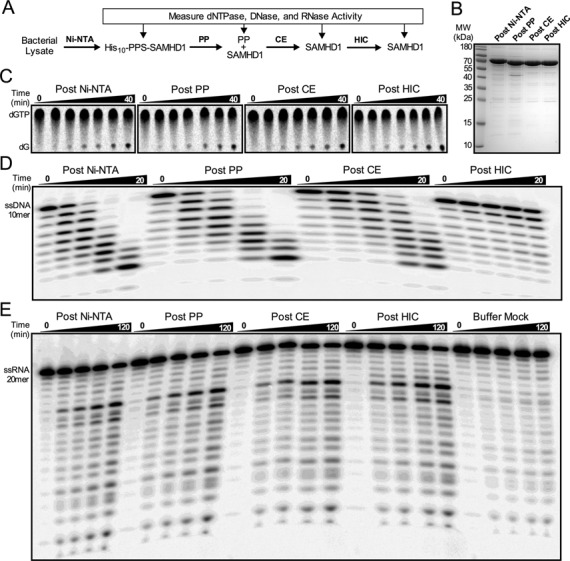
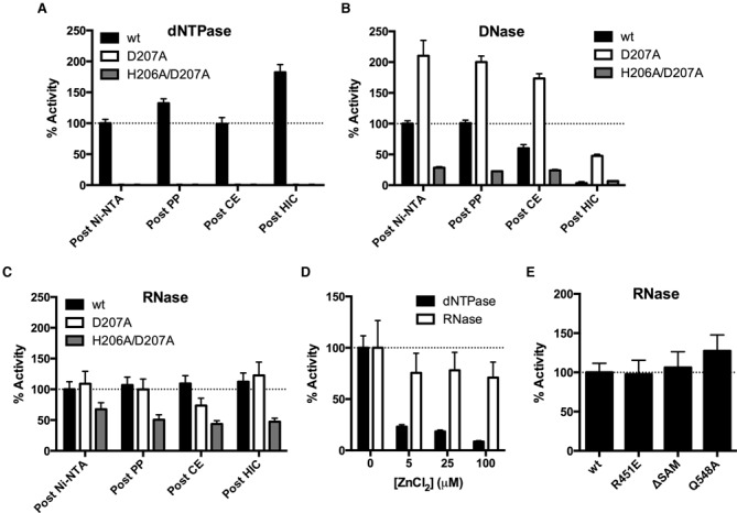
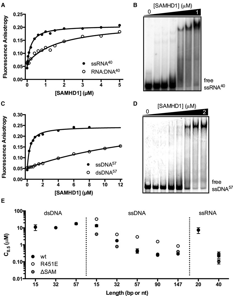
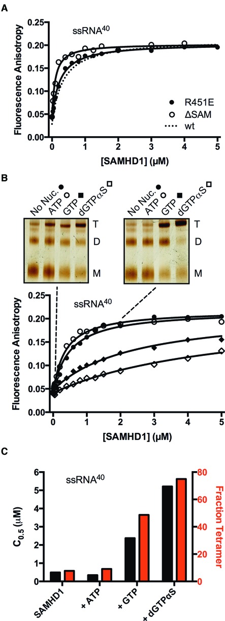
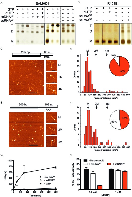
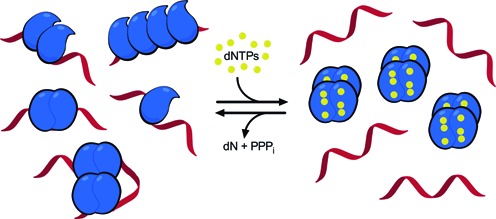
The HIV-1 restriction factor SAMHD1 is a tetrameric enzyme activated by guanine nucleotides with dNTP triphosphate hydrolase activity (dNTPase). In addition to this established activity, there have been a series of conflicting reports as to whether the enzyme also possesses single-stranded DNA and/or RNA 3'-5' exonuclease activity. SAMHD1 was purified using three chromatography steps, over which the DNase activity was largely separated from the dNTPase activity, but the RNase activity persisted. Surprisingly, we found that catalytic and nucleotide activator site mutants of SAMHD1 with no dNTPase activity retained the exonuclease activities. Thus, the exonuclease activity cannot be associated with any known dNTP binding site. Monomeric SAMHD1 was found to bind preferentially to single-stranded RNA, while the tetrameric form required for dNTPase action bound weakly. ssRNA binding, but not ssDNA, induces higher-order oligomeric states that are distinct from the tetrameric form that binds dNTPs. We conclude that the trace exonuclease activities detected in SAMHD1 preparations arise from persistent contaminants that co-purify with SAMHD1 and not from the HD active site. An in vivo model is suggested where SAMHD1 alternates between the mutually exclusive functions of ssRNA binding and dNTP hydrolysis depending on dNTP pool levels and the presence of viral ssRNA.


Figures

Figure 1.
Figure 1.
Copurification analysis of the dNTPase, DNase, and RNase activities. (A) SAMHD1 was purified by the indicated chromatography steps, with the three enzymatic activities tested at each point. (B) SDS-PAGE analysis of the purified SAMHD1 proteins (6 μg) with detection by Coomassie Brilliant Blue. Representative time course data is shown for the (C) RP-TLC dNTPase assay of SAMHD1 (0.5 μM) with dGTP (1 mM), (D) the PAGE ssDNase assay of SAMHD1 (0.5 μM) and ssDNA10 (1 μM), and (E) the PAGE ssRNase activity of SAMHD1 (0.5 μM) and ssRNA20 (1 μM).
Figure 2.
Figure 2.
The exonuclease activities cannot be attributed to the SAMHD1 active site. (A) dNTPase activities of wild-type and mutant enzymes were measured after each purification step. The D207A and H206A/D207A enzymes had no detectable dNTPase activities. (B) ssDNA exonuclease activities and (C) ssRNA exonuclease activities after each purification step. Each wild-type and mutant enzyme activity was normalized to the corresponding activity of the wild-type enzyme measured after the Ni-NTA purification step. (D) Rates of dNTPase or ssRNA exonuclease activity were measured with the indicated concentration of ZnCl2 (in the presence of 5 mM MgCl2). (E) The normalized ssRNA exonuclease activities of the wild-type, R451E, ΔSAM or Q548A enzymes. All activities were measured after the CE chromatography step (Figure 1A).
Figure 3.
Figure 3.
SAMHD1 is a single-stranded nucleic acid binding protein. (A) Binding of SAMHD1 to a 40mer 5′ FAM-labeled ssRNA (10 nM) and a 40 bp RNA:DNA hybrid duplex (10 nM) were measured by following the increase in fluorescence anisotropy of the FAM fluorophore upon nucleic acid binding. (B) Electrophoretic mobility shift (EMSA) measurements of SAMHD1 binding to ssRNA40 (10 nM). (C) The binding of SAMHD1 to a 57mer 5′ FAM-labeled ssDNA (50 nM) and a 57 bp DNA duplex (50 nM) were measured by following the increase in fluorescence anisotropy of the FAM fluorophore upon nucleic acid binding. (D) EMSA measurements of SAMHD1 binding to ssDNA57 (50 nM).
Figure 4.
Figure 4.
Binding of SAMHD1 to ssRNA40 does not require the SAM domain and is inhibited by guanine nucleotide-induced protein tetramerization. (A) Fluorescence anisotropy changes accompanying binding of the ΔSAM deletion mutant and the monomeric R451E mutant to ssRNA40 (10 nM). The binding curve for wt-SAMHD1 was very similar under the same conditions and is shown for comparison (dashed line). (B) Binding of wt-SAMHD1 to ssRNA40 was measured in the absence of nucleotide, and in the presence of ATP (non-activator), GTP, or dGTPαS (all at 1 mM concentration). For each nucleotide condition, the oligomeric state of SAMHD1 at two concentrations is indicated with the dashed lines (0.1 and 2 μM), which was determined by glutaraldehyde crosslinking and SDS-PAGE (insets). (C) The C0.5 values (black bars) and fraction of total SAMHD1 in the tetrameric form (red bars) for each nucleotide condition are correlated.
Figure 5.
Figure 5.
Longer ssRNA, but not ssDNA, induces higher oligomeric forms of SAMHD1 that are distinct from the tetramers induced by dNTP activation. (A) wt-SAMHD1 (1 μM) was crosslinked with glutaraldehyde in the presence of the indicated combinations of GTP (5 mM), dUTP (1 mM), ssDNA90 (2.5 μM), or ssRNA90 (2.5 μM) and the oligomeric forms were resolved by SDS-PAGE. Under conditions of this assay <10% of the dUTP was hydrolyzed by SAMHD1 and <5% of the ssRNA or ssDNA was hydrolyzed by the trace exonuclease activities that were present. (B) Glutaraldehyde crosslinking of the R451E mutant under identical conditions as wt-SAMHD1 showed that ssDNA, ssRNA and nucleotides were incapable of inducing higher order oligomeric forms. (C) AFM images of the wt-SAMHD1 complexes formed on the 60 nt ssDNA overhang of the 295 bp duplex AFM DNA construct (depicted) at 10:1 ratio. Bar size 200 nm. Shown to the right are representative images of complexes with protein volumes corresponding to SAMHD1 monomer (M), two monomers (2M) and four monomers (4M). Bar size 50 nm. (D) A histogram of the measured volumes of the globular ssDNA-protein complexes shows a distribution that is dominated by monomer complexes. The relative proportion of globular (red) and extended (white) protein complexes are shown in the inset pie chart. (E) AFM images of the wt-SAMHD1 complexes formed on the 102 nt ssRNA overhang of the 200 bp hybrid RNA:DNA duplex AFM construct (depicted) at 10:1 ratio. Bar size is 200 nm. On the right are representative images of complexes corresponding to SAMHD1 monomer (M), two monomers (2M) and four monomers (4M). Bar size is 50 nm. Complexes with protein volumes corresponding to monomer, dimer, and tetramer were observed in significant numbers. (F) A histogram of the measured volumes of the globular ssRNA-protein complexes showed a multimodal distribution with a shift towards higher order oligomeric species as compared to ssDNA. The relative proportion of globular (red) and extended (white) protein complexes are shown in the inset pie chart. (G) Despite inducing higher oligomeric forms of SAMHD1, ssRNA90 (2.5 μM) does not activate SAMHD1 (0.5 μM) for hydrolysis of the non-activating substrate dUTP. In contrast, addition of the activator GTP (0.1 mM) strongly induces dUTP hydrolysis. (H) SAMHD1 (0.5 μM) hydrolysis of low concentrations of the self-activating substrate dGTP (0.1 mM) is potently inhibited by ssRNA90 (2.5 μM) and to a lesser extent ssDNA90 (2.5 μM). Inhibition is not observed using high concentrations of dGTP (1 mM).
Figure 6.
Figure 6.
Possible binding modes of SAMHD1 to ssRNA and a model for the regulation of SAMHD1 dNTPase and RNA binding activities. In the low dNTP environment of resting immune cells, SAMHD1 exists in monomeric and dimeric forms that have high affinity for single-stranded RNA but possess no dNTPase activity. The figure depicts possible structures for RNA complexes with one, two and four monomers of SAMHD1 that are consistent with the biochemical, mutagenesis and AFM findings. The binding footprint for two adjacent monomers is ∼60 nt (Supplementary Figure S14), and two hypothetical binding modes for four monomers are depicted, consistent with chemical crosslinking (Figure 5A) and the elongated or globular complexes observed in AFM images (Figure 5E). Complexes of these types could form on HIV or LINE-1 ssRNA genomes. When dNTP levels increase, SAMHD1 can shift to its tetrameric form with high dNTPase activity but little affinity for single-stranded nucleic acids. Upon hydrolysis of available dNTP substrates by the activated SAMHD1 tetramers, the system will revert to the monomeric and dimeric states that are competent for ssRNA binding.

References

    1. Hult K., Berglund P. Enzyme promiscuity: mechanism and applications. Trends Biotechnol. 2007;25:231–238. - PubMed
    1. Pandya C., Farelli J.D., Dunaway-Mariano D., Allen K.N. Enzyme promiscuity: engine of evolutionary innovation. J. Biol. Chem. 2014;289:30229–30236. - PMC - PubMed
    1. Richardson C.C., Kornberg A. A deoxyribonucleic acid phosphatase-exonuclease from escherichia coli. I. Purification of the enzyme and characterization of the phosphatase activity. J. Biol. Chem. 1964;239:242–250. - PubMed
    1. Suck D. DNA recognition by DNase I. J. Mol. Recognit. 1994;7:65–70. - PubMed
    1. Stivers J.T., Nagarajan R. Probing enzyme phosphoester interactions by combining mutagenesis and chemical modification of phosphate ester oxygens. Chem. Rev. 2006;106:3443–3467. - PMC - PubMed

Publication types

MeSH terms