. 2015 Jul 27;43(13):6631-48.
doi: 10.1093/nar/gkv612. Epub 2015 Jun 22.

Post-translational environmental switch of RadA activity by extein-intein interactions in protein splicing

Affiliations

Post-translational environmental switch of RadA activity by extein-intein interactions in protein splicing

Natalya I Topilina et al. Nucleic Acids Res. .

Abstract

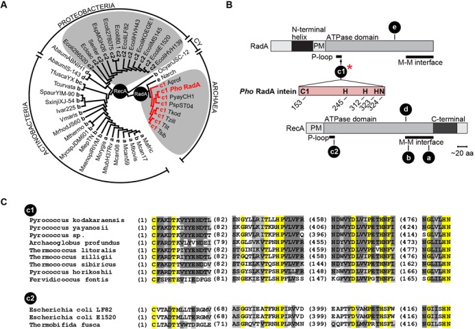
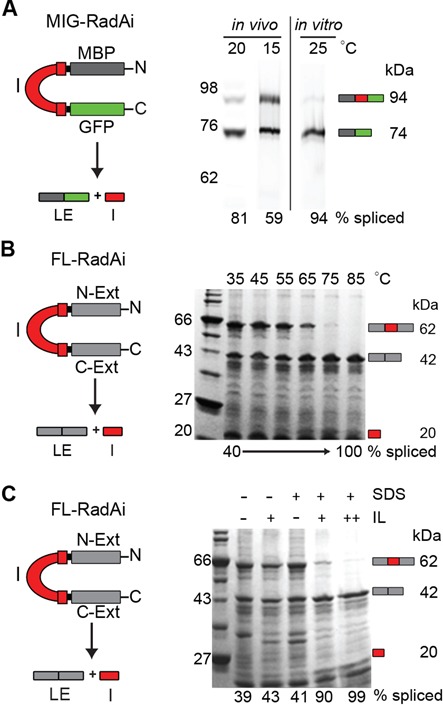
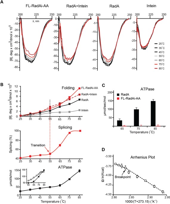
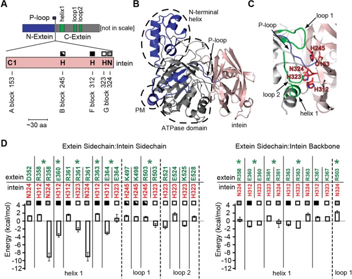
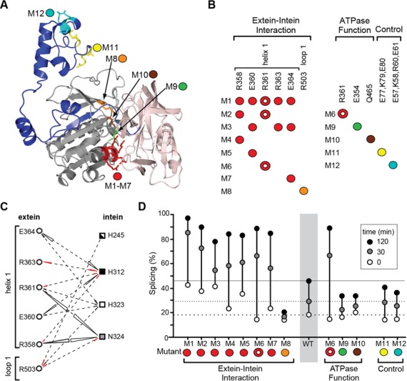
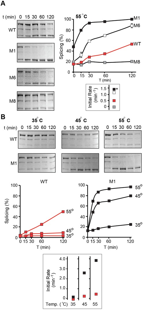
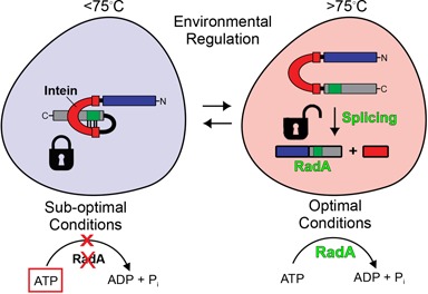
Post-translational control based on an environmentally sensitive intervening intein sequence is described. Inteins are invasive genetic elements that self-splice at the protein level from the flanking host protein, the exteins. Here we show in Escherichia coli and in vitro that splicing of the RadA intein located in the ATPase domain of the hyperthermophilic archaeon Pyrococcus horikoshii is strongly regulated by the native exteins, which lock the intein in an inactive state. High temperature or solution conditions can unlock the intein for full activity, as can remote extein point mutations. Notably, this splicing trap occurs through interactions between distant residues in the native exteins and the intein, in three-dimensional space. The exteins might thereby serve as an environmental sensor, releasing the intein for full activity only at optimal growth conditions for the native organism, while sparing ATP consumption under conditions of cold-shock. This partnership between the intein and its exteins, which implies coevolution of the parasitic intein and its host protein may provide a novel means of post-translational control.


Figures

Figure 1.
Figure 1.
Distribution and diversity of RadA/RecA inteins among archaea and bacteria. (A) Neighbor-Joining tree. The tree was constructed based on the multiple alignment of 42 RadA/RecA extein amino acid sequences from archaea and bacteria (proteobacteria, actinobacteria and cyanobacteria—CY). The insertion points (a–e) are indicated together with abbreviated species names, listed in full in Table 1. Letters a–e reflect the chronology of intein discovery (54,55). The species containing an intein in insertion point c (either c1 or c2) are on a gray background. The precursor of the protein of interest, Pho RadA (bold, red), carries an intein in insertion point c, subpopulation c1. (B) Intein insertion points. Intein locations are shown along RadA (c1 and e) and RecA (a–d) relative to structural and functional domains. The intein of interest, Pho RadA, is in insertion point c1 (red asterisk), which is located at the end of the RadA P-loop in the ATPase domain. Conserved, catalytically important amino acid residues and their positions are shown in single-letter code in the intein, as further identified in Figure 4A. Abbreviations: PM, polymerization motif; M–M interface, monomer–monomer interface. (C) Multiple sequence alignment of the inteins (splicing domains) from insertion point c. Comparative analysis of the splicing domains from inteins occupying insertion point c in RadA and RecA proteins revealed substantial differences in amino acid sequences between archaeal (insertion c1) and bacterial (insertion c2) inteins indicating independent acquisition of inteins by RadA and RecA in insertion point c.
Figure 2.
Figure 2.
RadA intein splicing is regulated by its native exteins in temperature and solution-dependent manner. (A) RadA intein splicing in foreign exteins is efficient even at low temperatures. The RadA intein was cloned into the non-native extein context (pMIG-RadAi; Table 2) and the recombinant protein was overexpressed in BL21 Star (DE3). The MIG-RadAi precursor (left) consists of the RadA intein (I; red) with short native exteins fused to MBP (dark gray) and GFP (green). The precursor (94 kDa) and the ligated exteins (LE; 74 kDa) were visualized by in-gel GFP fluorescence. More than 50% of precursor was spliced in vivo at 15°C and >80% was spliced at 20°C. The MIG-RadAi precursor recovered from a 15°C induction spliced efficiently (94% splicing) in vitro at 25°C within 1 h. (B) RadA intein splicing in the native exteins is inefficient at low temperatures, but efficient at high temperature. In the FL-RadAi precursor (left) the RadA intein (red) is flanked by its native exteins (N-Ext and C-Ext; gray). RadA intein splicing of the Ni-NTA purified FL-RadAi precursor was visualized in a Coomassie blue-stained gel. Accumulation of the spliced intein (I; 20 kDa) and the ligated exteins (LE; 42 kDa) and disappearance of the FL-RadAi precursor (62 kDa) were observed at high temperatures. A plot of the data from Figure 2B is shown in Figure 3B (middle panel). (C) Solution effects on RadA intein splicing in the native exteins. RadA intein splicing of FL-RadAi is shown in the presence 1.25% (+) and 2.5% (++) of ionic liquid (IL) 1-butyl-3-methylimidazolium chloride and 0.5% (+) SDS for samples incubated at 25°C for 30 min.
Figure 3.
Figure 3.
Temperature-dependent structure transition and ATPase activity of the Pho RadA protein and the precursor. (A) Far UV circular dichroism (CD) spectra. CD spectra of the splicing-inactive FL-RadAi-AA precursor, the RadA protein, RadA + Intein and Intein alone were recorded from 25–85°C with 10°C increments. (B) Temperature dependence of secondary structure transitions, splicing and ATPase activity. Top: Ellipticity at 217–223 nm measured for the four protein combinations in panel A. Middle: FL-RadAi splicing plot derived from data in Figure 2B. Splicing increases sharply at the structural transition, above 55°C. Bottom: ATPase activity of RadA. Inset corresponds to data used in panel D. CD data are from the experiment represented in panel A. Splicing and ATPase activity measurements in all panels were performed in triplicate at the temperatures indicated and error bars represent standard deviation. (C) ATPase activity of RadA and FL-RadAi at different temperatures. ATPase activity of inteinless RadA (black) increased with temperature while FL-RadAi (red) containing the inactive intein had no detectable ATPase activity. The experiment was performed in triplicate and error bars represent standard deviation. (D) Pho RadA exhibits biphasic ATPase activity. The Arrhenius plot demonstrates two distinct catalytic modes (55–76 and 76–83°C) with a slope change (Breakpoint) close to 76°C. The experiment was performed in triplicate and error bars represent standard deviation.
Figure 4.
Figure 4.
Model of interactions between the RadA exteins and the intein. (A) Linear representation of the RadA exteins and the intein. The intein (pink) is in the P-loop of the RadA ATPase domain. The N-extein and the C-extein are shown in blue and gray, respectively, as in panel B and C. Computational analysis predicted that residues within helix 1, loop 1 and loop 2 (green), located in the C-extein, interact strongly with the intein catalytic site. The catalytic residues of the intein are indicated by the one-letter amino acid code and their positions and conserved sequence blocks are shown. These residues, H245, H312, H323 and N324 are marked by different squares also used in panel D and Figure 5C. (B) Putative 3D structure of the Pho FL-RadAi precursor was generated by computer modeling based on avaliable structures of the Pho RadA intein (PDB ID: 4E2T) and Sulfolobus solfatarius RadA protein (PDB ID: 2ZUB). The large contact surface can be seen between the intein (pink) and C-extein (gray). Functionally important domains of the RadA protein: N-terminal helix, polymerization domain (PM) and ATPase domain, are demarcated by dashed lines. (C) The intein active site is at the extein–intein interface and has contacts with the C-extein. Catalytically important residues of the intein (pink) C153, H245, H312, H323 and N324 are shown as red sticks alongside the C-extein (gray). Computational analysis predicted that residues within helix 1, loop 1 and loop 2 (all green) in the C-extein interact strongly with the catalytic residues of the intein. (D) Calculated interaction energies. Interaction energies between the extein residues (green) and the intein active-site residues (red) are shown. Only amino acid residue pairs with interaction energy values >1 kcal/mol are shown. ‘Extein Sidechain:Intein Sidechain’ (left) and ‘Extein Sidechain:Intein Backbone’ (right) interactions are plotted separately. The intein catalytic amino acid residues (H245, H312, H323 and N324) are marked by squares as in panel A. The error bars in the figure are the standard deviations obtained from energy calculations for 10 modeled precursor structures. The extein amino acid residues which show the strongest interactions with the intein (green asterisks) were chosen for experimental studies of the extein–intein interactions.
Figure 5.
Figure 5.
The extein–intein interactions have a profound effect on the intein splicing. (A) Locations of the extein mutations. Mutations M1–M12 are highlighted on the structure model of the Pho FL-RadAi precursor. (B) Mutant design. The extein–intein interaction mutants (M1–M8) were designed based on the interaction energy analysis (Figures 4D and 5C); the ATPase function mutants (M6, M9, M10) carry mutations which disrupt ATPase activity of the RadA protein; control mutants (M11 and M12) carry mutations at amino acid residues that are distant from the intein and are unlikely to interact with the intein active site. R361 in M6 (circle with white center) is a residue that emerged from the computational screen as an interacting residue and is also a catalytic residue of the RadA protein. The color-coding corresponds to the model in (A). (C) The extein–intein interactions. The extein amino acid residues selected for experimental studies (derived from Figure 4D, asterisks) are shown with interacting intein residues by dashed lines. The interaction energies between amino acid residue pairs are shown with triangles, the area which corresponds to the value of the interaction energy (negative, black; positive, red). (D) Characterization of the FL-RadAi extein mutants. Splicing was monitored in vitro at 55°C in comparison with the wild-type FL-RadAi precursor (WT). The percentages of splicing at three time points: 0, 30 and 120 min, are plotted. The 0 min time-point reflects in vivo splicing during induction at 46°C.
Figure 6.
Figure 6.
Extein–intein interactions affect temperature dependence of Pho RadA intein splicing. (A) Kinetics of splicing of extein mutants M1, M6 and M8. Mutants are compared with the wild-type FL-RadAi precursor (WT) at 55°C. Proteins were overexpressed in BL21 Star (DE3) cells at 46°C and Ni-NTA purified. The experiment was performed in triplicate and error bars represent standard deviation. All splicing assays have very small deviations, obscuring error bars in the plot. Initial rates of splicing are shown on the right. * indicates that the initial rate for M1 was calculated for the sample that had 50% of the precursor converted in vivo and therefore is likely to be an underestimate. (B) Splicing of extein mutant M1. WT and M1 mutant proteins were overexpressed in ArcticExpress (DE3) cells at 12°C. Mutant M1 and the wild-type FL-RadAi precursor (WT) were compared at 55, 45 and 35°C. All splicing assays have very small deviations, obscuring error bars in the plot. The experiment was performed in triplicate and error bars represent standard deviation. Initial rates of splicing at different temperatures are shown on the right.
Figure 7.
Figure 7.
Extein–intein partnership regulates splicing. At suboptimal conditions of temperature and solution environment, the exteins place a lock on splicing through interactions with the intein in 3D space and ATP (boxed) accumulates (left). Under optimal conditions of high temperature and/or solution environment (right) the lock imposed by extein–intein interactions is released and splicing ensues to yield an active RadA protein (green).

Similar articles

Cited by

References

    1. Novikova O., Topilina N., Belfort M. Enigmatic distribution, evolution, and function of inteins. J. Biol. Chem. 2014;289:14490–14497. - PMC - PubMed
    1. Liu X.Q. Protein-splicing intein: genetic mobility, origin, and evolution. Annu. Rev. Genet. 2000;34:61–76. - PubMed
    1. Gogarten J.P., Senejani A.G., Zhaxybayeva O., Olendzenski L., Hilario E. Inteins: structure, function, and evolution. Annu. Rev. Microbiol. 2002;56:263–287. - PubMed
    1. Swithers K.S., Senejani A.G., Fournier G.P., Gogarten J.P. Conservation of intron and intein insertion sites: implications for life histories of parasitic genetic elements. BMC Evol. Biol. 2009;9:303. - PMC - PubMed
    1. Callahan B.P., Topilina N.I., Stanger M.J., Van Roey P., Belfort M. Structure of catalytically competent intein caught in a redox trap with functional and evolutionary implications. Nat. Struct. Mol. Biol. 2011;18:630–633. - PMC - PubMed

Publication types

LinkOut - more resources