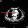
Squamous cell carcinoma of unknown origin metastasising to the right atrium causing acute heart failure
- PMID: 26101300
- PMCID: PMC4480105
- DOI: 10.1136/bcr-2015-210042
Squamous cell carcinoma of unknown origin metastasising to the right atrium causing acute heart failure
Abstract
We describe a case of metastasis to the heart, which was initially suspected to be a myxoma, causing acute right heart failure. Emergency surgery was carried out by opening the right atrium and superior vena cava, and debulking the tumour in a piecemeal fashion, providing temporary relief of symptoms. The histology showed this to be metastatic squamous cell carcinoma possibly of head and neck origin. This is extremely rare, with few published cases. Full endoscopic and CT, including positron emission tomography CT, investigation of the head and neck was performed with no primary findings. Only two such cases of squamous cell carcinoma of unknown origin metastasising to the heart have been described, and, in both cases, the patients died within several weeks of diagnosis. This patient remains alive 2 months postoperatively and is receiving radiotherapy to the chest, but his prognosis remains poor.
2015 BMJ Publishing Group Ltd.
Figures


References
-
- Ewer MS, Edward THY. Cancer and the heart. People's medical publishing house. 2nd edn Connecticut, USA: 2013.
-
- Columbus M. De re anatomica. Venice: N Bevilacque, 1559:14.
-
- Yater W. Tumors of the heart and pericardium: pathology, symptomatology and report of nine cases. Arch Intern Med 1931;48:627–66. 10.1001/archinte.1931.00150040093007 - DOI
-
- Barnes A, Beaver D, Snell AM. Primary sarcoma of the heart: report of a case with electrocardiographic and pathological studies. Am Heart J 2003;9:480–91. 10.1016/S0002-8703(34)90096-X - DOI
Publication types
MeSH terms
LinkOut - more resources
Full Text Sources
Other Literature Sources
Medical
