

doi: 10.3978/j.issn.2072-1439.2015.04.60.
Inflammatory myofibroblastic tumour of the lung: a reactive lesion or a true neoplasm?
Affiliations
- PMID: 26101648
- PMCID: PMC4454865
- DOI: 10.3978/j.issn.2072-1439.2015.04.60
Item in Clipboard
Inflammatory myofibroblastic tumour of the lung: a reactive lesion or a true neoplasm?
J Thorac Dis.
2015 May.
Abstract
Inflammatory myofibroblastic tumour (IMT) of the lung represents an extremely rare type of inflammatory pseudo tumor that appears most commonly in children and young individuals. There has been an ongoing controversy whether an IMT is a reactive lesion or a true neoplasm making the further management extremely challenging. Purpose of the paper is through a literature review to highlight the existence of this rare tumour along with its key features and the management options available.
Keywords: Lung neoplasms; inflammatory myofibroblastic tumors (IMTs).
Figures
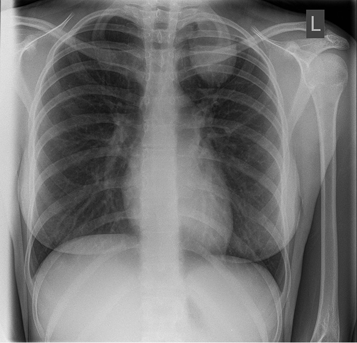
Chest X-ray (CXR) showing a solitary left upper lobe lung mass.

MRI scan demonstrating a lesion in the left upper lobe of the lung. MRI, magnetic resonance imaging.
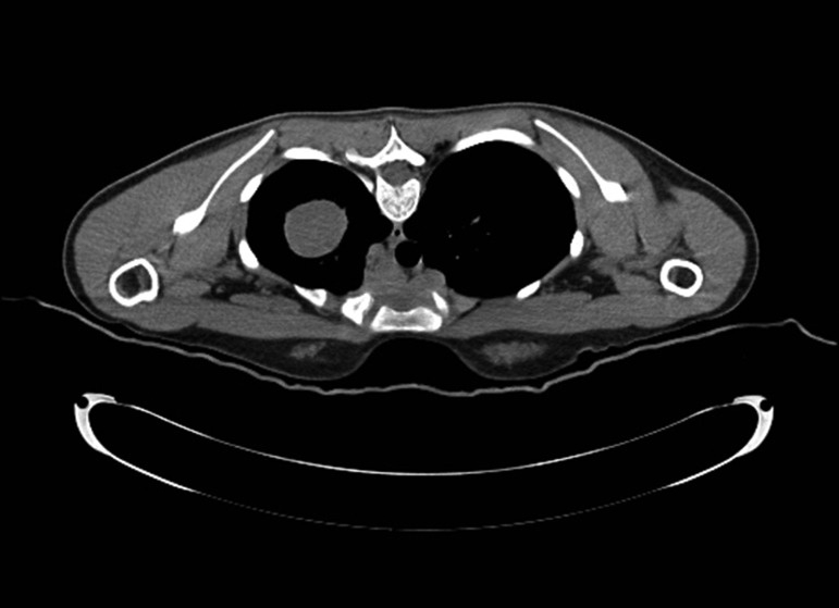
CT scan depicting a white colour mass in the left lung. CT, computed tomography.
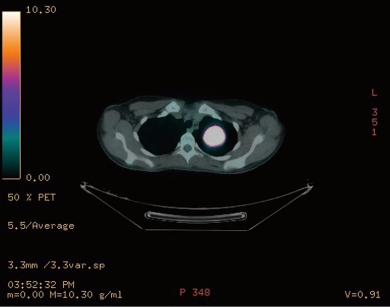
PET CT shows FDG avid lesion in the left upper lobe of the lung. CT, computed tomography; PET, positron emission tomography; FDG, fluorodeoxyglucose.
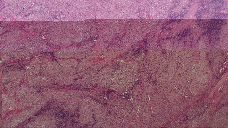
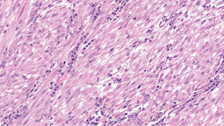
A low power image of the tumor which comprises interlacing fascicles of bland spindle cells which obliterate the underlying architecture of the lung (H&E ×20 magnification).
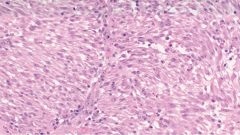
Spindle cell component that is admixed with inflammatory cells including plasma cells and eosinophils. The spindle cells show minimal pleomorphism and mitotic figures which are sparse (H&E ×200 magnification).
Spindle cell component that is admixed with inflammatory cells including plasma cells and eosinophils. The spindle cells show minimal pleomorphism and mitotic figures which are sparse (H&E ×200 magnification).
References
-
- Elmadi A, Rami M, Khattala K, et al. Pseudotumeur inflammatoire pulmonaire chez un enfant. J pediatr Puericulture 2011;24:69-71.
-
- Pinilla I, Herrero Y, Torres MI, et al. Tumor inflamatorio miofibroblástico pulmonar. Radiología 2007;49:53-5. - PubMed
-
- Matsubara O, Mark EJ, Ritter JH. Pseudoneoplastic lesions of the lungs, pleural surfaces, and mediastinum. In: Wick MR, Humphrey PA, Ritter JH. editors. Pathology of pseudoneoplastic lesions. New York: Lippincott-Raven, 1997:100-9.
-
- Cerfolio RJ, Allen MS, Nascimento AG, et al. Inflammatory pseudotumors of the lung. Ann Thorac Surg 1999;67:933-6. - PubMed
-
- Ishida T, Oka T, Nishino T, et al. Inflammatory pseudotumor of the lung in adults: radiographic and clinicopathological analysis. Ann Thorac Surg 1989;48:90-5. - PubMed
LinkOut - more resources
Full Text Sources