. 2015 Sep:63:158-67.
doi: 10.1016/j.biomaterials.2015.06.009. Epub 2015 Jun 11.

Enhanced therapeutic neovascularization by CD31-expressing cells and embryonic stem cell-derived endothelial cells engineered with chitosan hydrogel containing VEGF-releasing microtubes

Affiliations

Enhanced therapeutic neovascularization by CD31-expressing cells and embryonic stem cell-derived endothelial cells engineered with chitosan hydrogel containing VEGF-releasing microtubes

Sangho Lee et al. Biomaterials. 2015 Sep.

Abstract

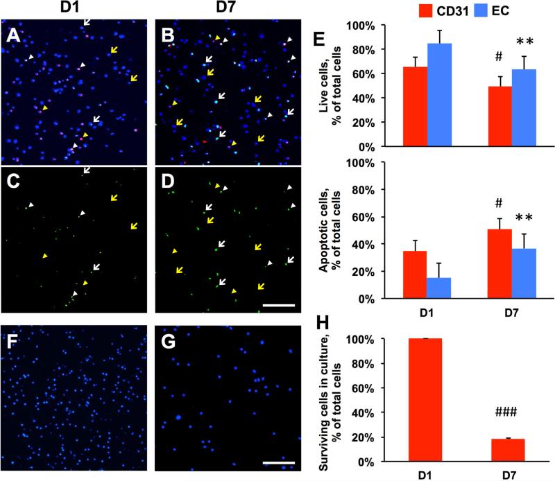
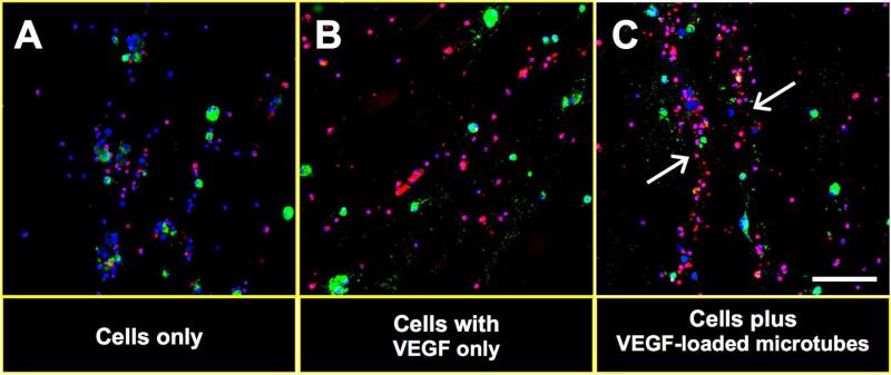
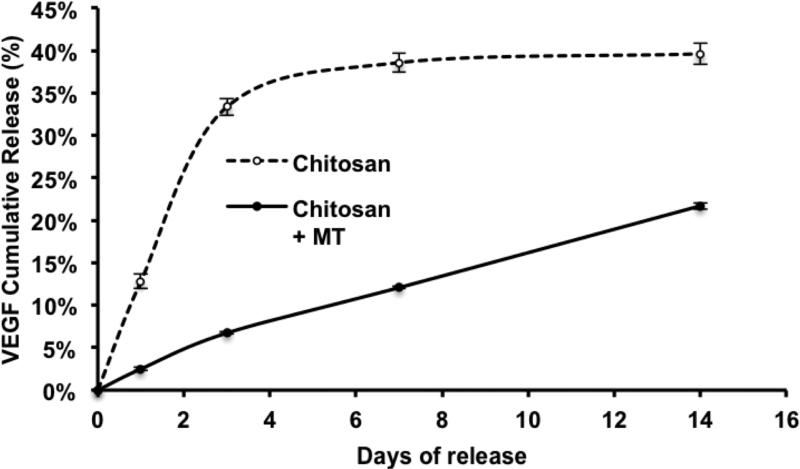
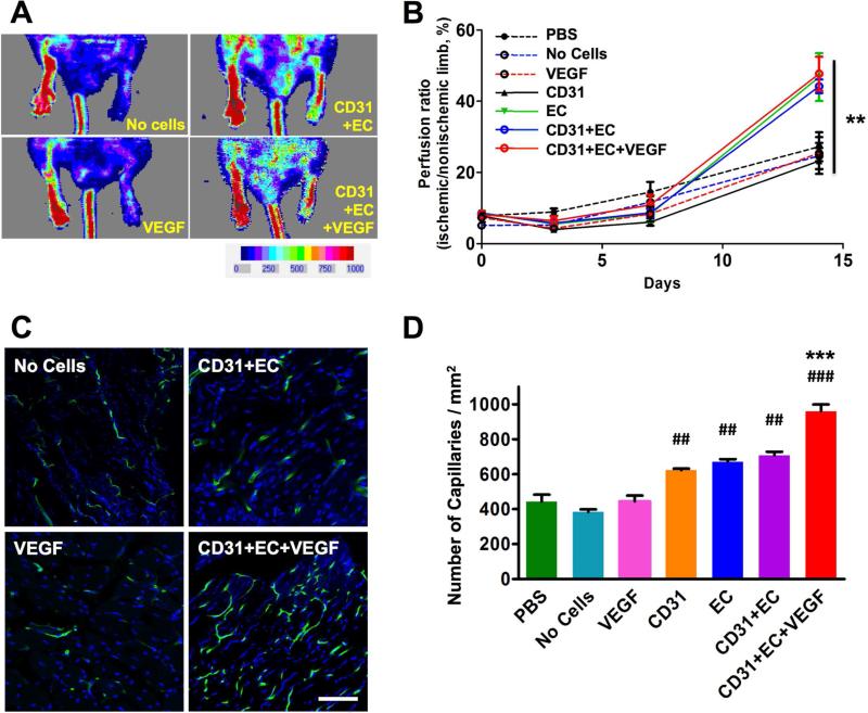
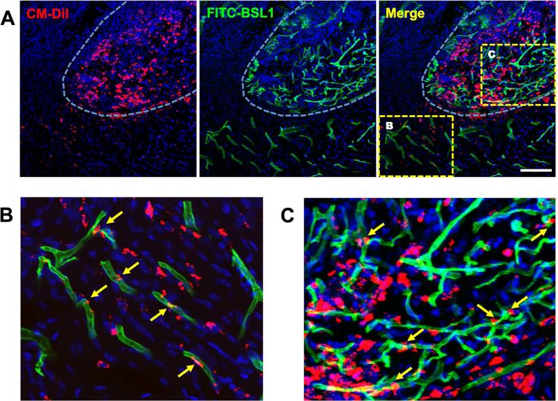
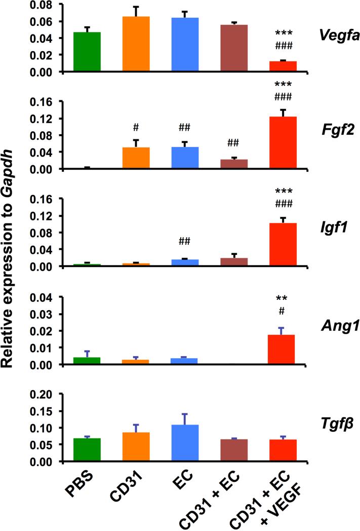
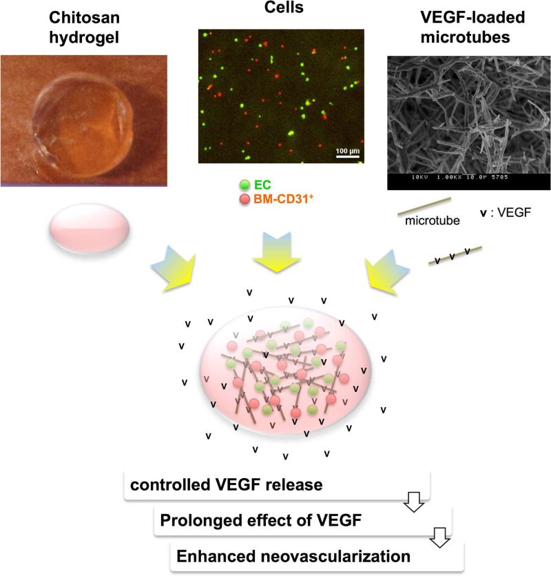
Various stem cells and their progeny have been used therapeutically for vascular regeneration. One of the major hurdles for cell-based therapy is low cell retention in vivo, and to improve cell survival several biomaterials have been used to encapsulate cells before transplantation. Vascular regeneration involves new blood vessel formation which consists of two processes, vasculogenesis and angiogenesis. While embryonic stem cell (ESC)-derived endothelial cells (ESC-ECs) have clearer vasculogenic potency, adult cells exert their effects mainly through paracrine angiogenic activities. While these two cells have seemingly complementary advantages, there have not been any studies to date combining these two cell types for vascular regeneration. We have developed a novel chitosan-based hydrogel construct that encapsulates both CD31-expressing BM-mononuclear cells (BM-CD31(+) cells) and ESC-ECs, and is loaded with VEGF-releasing microtubes. This cell construct showed high cell survival and minimal cytotoxicity in vitro. When implanted into a mouse model of hindlimb ischemia, it induced robust cell retention, neovascularization through vasculogenesis and angiogenesis, and efficiently induced recovery of blood flow in ischemic hindlimbs. This chitosan-based hydrogel encapsulating mixed adult and embryonic cell derivatives and containing VEGF can serve as a novel platform for treating various cardiovascular diseases.

Keywords: CD31(+) cells; Chitosan hydrogel; Embryonic stem cells; Lipid microtubes; Vascular regeneration.


Figures

Figure 1
Figure 1. Better survival of co-cultured mBM-CD31+ cells and mESC-ECs in chitosan hydrogel
A-D. TUNEL staining and confocal examination. mBM-CD31+ cells (CM-DiI labeled, red fluorescence) and mESC-ECs (non-labeled) were embedded in chitosan hydrogel and cultured. The hydrogel patches were collected at days 1 (D1, A and C) and 7 (D7, B and D) and were stained for DAPI (blue) and TUNEL (green). A and B are merged images of all three channels (DAPI, CM-DiI, TUNEL), whereas C and D show only TUNEL positive signals. Confocal microscopy examination showed live mBM-CD31+ cells (yellow arrowheads, red cells), apoptotic mBM-CD31+ cells (white arrowheads, green/red cells), live mESC-ECs (yellow arrows, blue nuclei), and apoptotic mESC-ECs (white arrows, green cells). E. Quantitative evaluation of the percent of live cells and apoptotic cells per total cells (live cells plus apoptotic cells). F-G. Cell survival in culture without chitosan hydrogel. F and G are representative images of DAPI-stained mBM-CD31+ cells in culture dishes at days 1 (F) and 7 (G). H. Quantitative evaluation of the percent of surviving cells at day 7 per cells at day 1. Scale bar: 100 μm. N = 3, ###P < 0.001, #P < 0.05 vs. D1 CD31, **P < 0.01 vs. D1 EC.
Figure 2
Figure 2. Tubular alignment of cell mixture in chitosan hydrogel after application of VEGF-loaded microtubes
mBM-CD31+ cells (CM-DiI labeled, red) and mESC-ECs (SP-DiO labeled, green) were embedded in chitosan hydrogel in the absence of VEGF165-loaded microtubes (A), presence of VEGF165 only (B), and presence of VEGF165-loaded microtubes (C). The hydrogel-cell mixture was collected at day 4 and stained with DAPI (blue). Confocal microscopic examination showed tube-like cellular arrangement (white arrows) only in the presence of VEGF165-loaded microtubes (C). Scale bar: 100 μm.
Figure 3
Figure 3. Release profiles of VEGF from lipid microtubes
The release kinetics of VEGF165 from chitosan hydrogel (Chitosan) containing bare VEGF165 or chitosan hydrogel containing VEGF165-loaded microtubes (Chitosan + MT) were examined. The cumulative release was calculated as the percentage of total amount released at the indicated time points divided by the initially loaded amount of VEGF165. Data were shown as means ± S.E.M.
Figure 4
Figure 4. Increase in blood flow and capillary density in ischemic hindlimbs treated with various cells and cell-chitosan patch
A. The blood perfusion was measured by LDPI at days 3, 7, and 14. The representative LDPI images at day 14 are shown. B. The quantitation of blood perfusion was plotted. N = 4 to 6, **P < 0.01. C. Representative images of capillaries in hindlimb. D. Quantitative analyses of capillary density at day 14. Scale bars: 100 μm. N = 5, ###P < 0.001 and ##P < 0.01 vs. PBS; ***P < 0.001 vs. CD31+EC. CD31: mBM-CD31+ cells, EC: mESC-EC, VEGF: VEGF165-loaded microtubes.
Figure 5
Figure 5. Incorporation of cells into vasculature
Confocal microscopic images of ischemic limb tissues transplanted with a chitosan patch encapsulating mBM-CD31+ cells and mESC-ECs with VEGF165-loaded microtubes were analyzed for the incorporation of cells into vasculature. A. All cells were labeled with CM-DiI (red). The mouse was perfused with FITC-BSL1 (green) to visualize the functional vessels at day 14. DAPI (blue). White dotted line marks transplanted chitosan hydrogel area which shows encapsulated cells (red) and invested blood vessels (green). B and C are magnified images of the boxed areas in A. B. Transplanted cells were migrated from hydrogel to the neighboring areas and were incorporated into the blood vessels as endothelial cells, showing vasculogenesis (yellow arrows). C. Transplanted cells in chitosan hydrogel area showed extensive vasculogenesis and angiogenesis by incorporation of transplanted cells (yellow arrows) into vessels and ingrowth of host vessels from surrounding vessels. Scale bar: 100 μm.
Figure 6
Figure 6. Increased mRNA expression of angiogenic factors in ischemic hindlimbs treated with chitosan-cell patch
The expression of Vegfa, Fgf2, Igf1, Ang1 and Tgfβ genes were examined by real-time RT-PCR of hindlimb tissues harvested at 4 weeks following HLI surgery. The expression level of each gene is plotted as a relative value to the expression level of Gapdh. N = 3. ###P < 0.001, ##P < 0.01, and #P < 0.05 vs. PBS; ***P < 0.001 and **P < 0.01 vs. CD31+EC.
Figure 7
Figure 7. Advantages of the lipid microtubes loaded with VEGF
The top portion shows photographs of a representative chitosan gel, ECs (green) and BM-CD31+ cells (red), and a high magnification (1000X) scanning electron micrograph of lipid microtubes. The bottom portion illustrates the combination of the three: cells (ECs as green dots and BM-CD31+ as red dots) and VEGF165 (v)-loaded microtubes (grey sticks) which are encapsulated by chitosan hydrogel (pink oval). The lipid microtubes provide controlled release of VEGF, prolonging the effects of VEGF and resulting in enhanced neovascularization.

Similar articles

Cited by

References

    1. Kinoshita M, Fujita Y, Katayama M, Baba R, Shibakawa M, Yoshikawa K, et al. Long-term clinical outcome after intramuscular transplantation of granulocyte colony stimulating factor-mobilized CD34 positive cells in patients with critical limb ischemia. Atherosclerosis. 2012;224(2):440–5. - PubMed
    1. Powell RJ. Update on clinical trials evaluating the effect of biologic therapy in patients with critical limb ischemia. Journal of vascular surgery. 2012;56(1):264–6. - PubMed
    1. Raval Z, Losordo DW. Cell therapy of peripheral arterial disease: from experimental findings to clinical trials. Circ Res. 2013;112(9):1288–302. - PMC - PubMed
    1. Carmeliet P, Jain RK. Molecular mechanisms and clinical applications of angiogenesis. Nature. 2011;473(7347):298–307. - PMC - PubMed
    1. Potente M, Gerhardt H, Carmeliet P. Basic and therapeutic aspects of angiogenesis. Cell. 2011;146(6):873–87. - PubMed

Publication types

MeSH terms

Substances