. 2015;14(16):2619-33.
doi: 10.1080/15384101.2015.1064565. Epub 2015 Jun 23.

Np9, a cellular protein of retroviral ancestry restricted to human, chimpanzee and gorilla, binds and regulates ubiquitin ligase MDM2

Affiliations

Np9, a cellular protein of retroviral ancestry restricted to human, chimpanzee and gorilla, binds and regulates ubiquitin ligase MDM2

Kristina Heyne et al. Cell Cycle. 2015.

Abstract

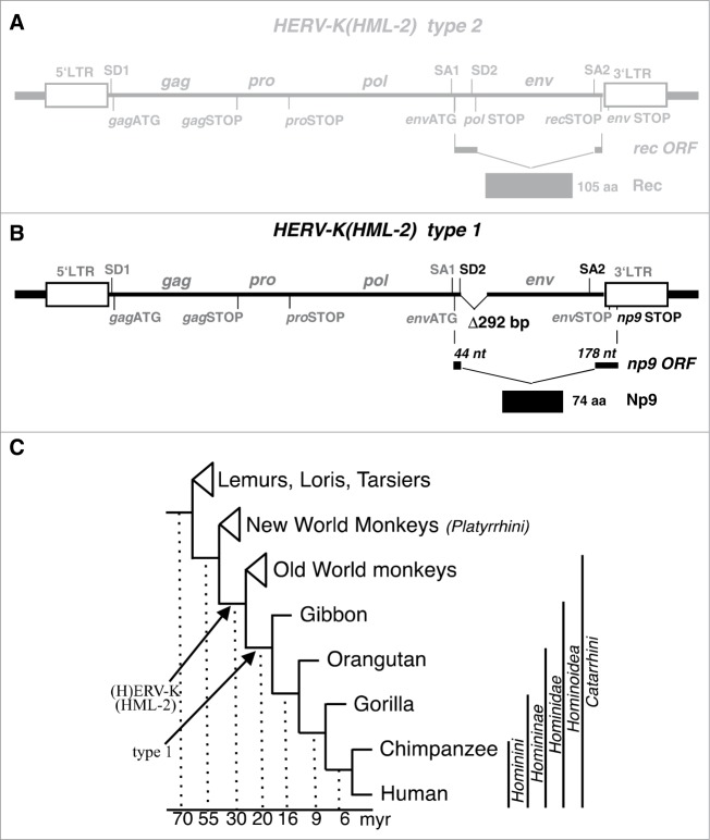
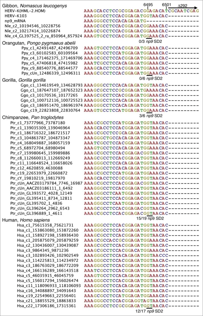
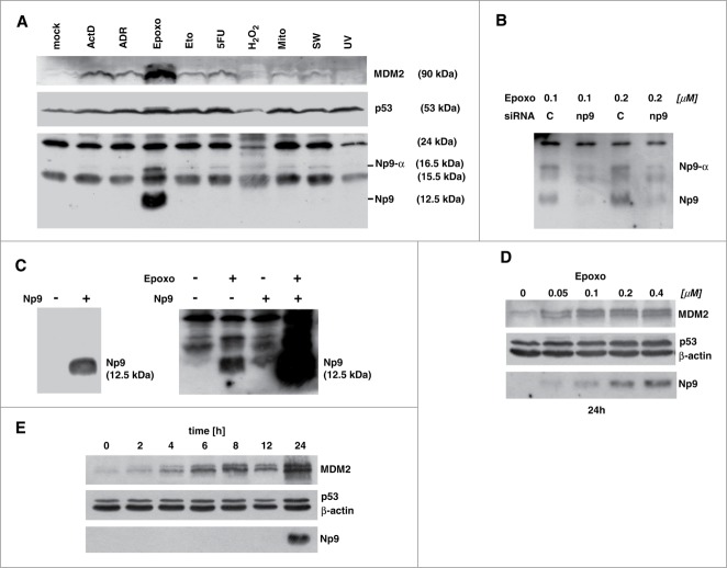
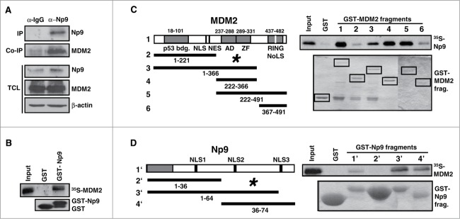
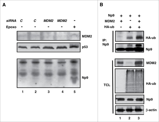
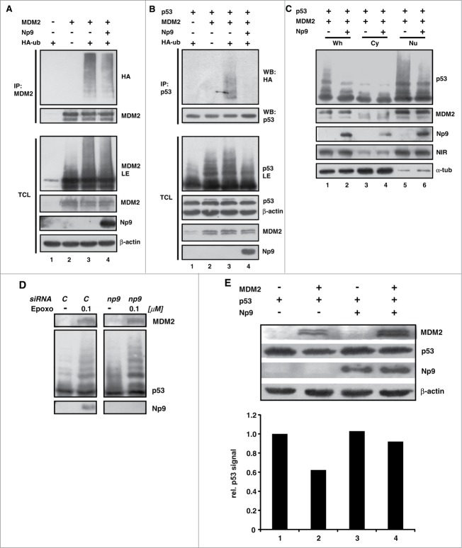
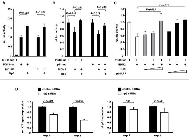
Humans and primates are long-lived animals with long reproductive phases. One factor that appears to contribute to longevity and fertility in humans, as well as to cancer-free survival, is the transcription factor and tumor suppressor p53, controlled by its main negative regulator MDM2. However, p53 and MDM2 homologs are found throughout the metazoan kingdom from Trichoplacidae to Hominidae. Therefore the question arises, if p53/MDM2 contributes to the shaping of primate features, then through which mechanisms. Previous findings have indicated that the appearances of novel p53-regulated genes and wild-type p53 variants during primate evolution are important in this context. Here, we report on another mechanism of potential relevance. Human endogenous retrovirus K subgroup HML-2 (HERV-K(HML-2)) type 1 proviral sequences were formed in the genomes of the predecessors of contemporary Hominoidea and can be identified in the genomes of Nomascus leucogenys (gibbon) up to Homo sapiens. We previously reported on an alternative splicing event in HERV-K(HML-2) type 1 proviruses that can give rise to nuclear protein of 9 kDa (Np9). We document here the evolution of Np9-coding capacity in human, chimpanzee and gorilla, and show that the C-terminal half of Np9 binds directly to MDM2, through a domain of MDM2 that is known to be contacted by various cellular proteins in response to stress. Np9 can inhibit the MDM2 ubiquitin ligase activity toward p53 in the cell nucleus, and can support the transactivation of genes by p53. Our findings point to the possibility that endogenous retrovirus protein Np9 contributes to the regulation of the p53-MDM2 pathway specifically in humans, chimpanzees and gorillas.

Keywords: MDM2; Np9; endogenous retrovirus; evolution; p53; ubiquitylation.


Figures

Figure 1.
Figure 1.
Provirus structure of HERV-K(HML-2) and germ line integration during primate evolution. (A) Structure of the more ancient HERV-K(HML) type 2 provirus harbouring the rec open reading frame. Thick lines at each end indicate host cell genomic DNA. LTR, Long-terminal Repeat. SD, SA, splice donor and splice acceptor sites. gag, pro, pol, env, group specific antigen, protease, polymerase and envelope protein open reading frames. nt, nucleotides. aa, amino acid residues. (B) Structure of HERV-K(HML-2) type 1 provirus. A 292 bp deletion at the 5′-end of the env open reading frame defines type 1 proviruses. Another mutation (see Figure 2) generated a new splice donor site (SD; highlighted in black), resulting in an alternatively spliced mRNA with the ORF encoding the 74 aa residue Np9 protein (for further details, see main text). (C) Evolutionary relationships among groups of primates and selected primate species (adapted from66). Arrows depict the fixation of HERV-K(HML-2) type 1 and type 2 proviruses. myr, million years.
Figure 2.
Figure 2.
Evolution in Hominoidea of the np9 splice donor site 2 (SD2) crucial for Np9 protein expression. A subregion of HERV-K(HML-2) type 1 homologous sequences identified in various Hominoidea genome sequences is shown as a multiple alignment. For comparisons, corresponding regions of reference sequences HERV-K(HML-2.HOM) (type 2) (Genbank acc. no. AF074086; 15), the previously identified HERV-K103 provirus (type 1) (AF164611; 14) and a previously described np9 mRNA sequence. The 3′ end of the np9 exon 2 is located at nt 6495 and the 292 bp deletion, defining type 1 proviruses, starts at nt 6502 with respect to the HERV-K(HML-2.HOM) sequence. Approximate locations of HERV-K(HML-2) homologous type 1 sequences in the various primate genomes (see Materials and Methods) are indicated by sequence names left from actual sequences as ‘species abbreviation_chromosome no._start position_end position’. The critical GA–GT mutation generating the 5′ end of np9 intron 2 is indicated by a horizontal line and the number of sequences displaying GT, relative to the total number of type 1 provirus sequences, is given below each of the multiple alignments.
Figure 3.
Figure 3.
Expression of Np9 protein in response to various stresses. (A) Proteasome inhibition causes accumulation of Np9. Exponentially growing Tera-1 cultures were either mock-treated or exposed to actinomycin D (ActD; 10 nM), adriamycin (ADR; 0.34 μM), epoxomicin (Epoxo; 0.4 μM), etoposide (Eto, 10 μM), 5-fluorouracil (5-FU; 375 μM), H2O2 (0.4 mM); mitomycin C (Mito; 3 μM); serum withdrawal (SW) and UV light (200 J/m2). Total protein extracts were prepared after 24 h, and 15 μg protein per lane was analyzed by western blotting, using monoclonal anti-MDM2 antibody 3G9 (1:2,000), monoclonal anti-p53 antibody DO-1 (1 : 2,000), and rat monoclonal anti-Np9 antibody 22E4 (1:5). (B) np9 transcripts sensitive to np9-specific siRNA produce 12.5 kDa and a 16.5 kDa Np9 proteins. Tera-1 cells were transfected for 24 h with scrambled siRNA (C=control; 20 nM) or a mixture of 4 np9 siRNAs (5 nM each). After transfection, the cells were treated with 2 concentrations of epoxomicin for another 24 h. Western blot analysis was as in (A). (C) H1299 cells negative for np9 give rise to a single signal of 12.5 kDa upon transfection for 24 h with plasmid pCMV-np9 containing the np9 ‘reference’ ORF (left panel). Tera-1 cells transfected with pCMV-np9 produce an Np9 signal of similar size as the endogenous Np9 produced under epoxomicin, and this signal is further increased in response to epoxomicin (right panel). Western blots were performed as in (A). (D) Dose-dependent increase in Tera-1 cells of Np9 expression after 24 h of treatment with increasing, sub-micromolar concentrations of epoxomicin. Western blot was performed as in A; β-actin was detected with monoclonal anti-β-actin antibody (1 : 10,000). (E) Late accumulation of Np9 in Tera-1 cells compared to MDM2 and p53, various times after the exposure to epoxomicin (0.4 μM).
Figure 4.
Figure 4.
Np9 associates with MDM2. (A) Np9 coprecipitates MDM2 from transfected H1299 cell extracts. Cells were transfected with expression plasmids pCMV-np9 and pcmdm2. At 24 h after transfection, cell extracts were incubated with irrelevant IgG antibody or with anti-Np9 antibody 22E4 (2 μg/IP). Precipitates were analyzed by western blotting. Proteins were detected as specified in the legend of Figure 2. IP, immunoprecipitation. Co-IP, co-immunoprecipitation. TCL, total cell lysate. (B) GST pull-down analysis. Lower panel: Equal load of GST and GST-Np9 on beads. Upper panel: In vitro translated 35S-labeled full-length human MDM2 was retained by GST-Np9 but not GST alone. Input shows 10 % of total 35S-MDM2. (C) Radiolabeled Np9 binds to the central acidic and zinc finger domain of MDM2 in GST pull-down assays. (D) Radiolabeled MDM2 associates with the C-terminal half of Np9. The indicated MDM2 fragments (1–6) and Np9 fragments (1′–4′) were fused to GST and incubated with in vitro translated 35S-Np9 or 35S-MDM2. While labeled protein failed to bind to GST alone, 35S-Np9 was retained primarily by GST-MDM2 fragments 3–5, and 35S-MDM2 was retained mostly by GST-Np9 fragments 3 and 4. Asterisks mark main interaction domains. p53-bdg., p53 binding domain. NLS, nuclear localization signal. NES, nuclear export signal. AD, acidic domain. ZF, zinc finger domain. RING is the domain important for ubiquitin ligase activity, intra/inter-molecular protein interactions and interaction with RNA. NoLS, nucleolar localization domain. Numbers denote amino acid residues.
Figure 5.
Figure 5.
Effect of MDM2 on the expression level and modification of Np9. (A) MDM2 knockdown increases p53 level but fails to cause accumulation of Np9. Proteasome inhibition increases the expression of Np9 to higher levels when MDM2 has been knocked down. Tera-1 cells were transfected with control siRNA (20 nM; C) or MDM2 siRNA (20 nM) for 24 h. Mock-transfected Tera-1 cultures treated with epoxomicin (100 nM) for 24 h served as positive control for Np9 expression. Western blot analysis was performed as detailed in the legend of Figure 2. (B) In vivo HA-ubiquitylation of Np9 in the presence of ectopic MDM2. H1299 cells harbouring low levels of MDM2 were transfected with pCMV-np9, pcmdm2 and pCMV-HA-ub producing HA-tagged ubiquitin, as indicated. At 24 h after transfection, proteasomal degradation was inhibited by MG132 (10 μM) for another 4 h. Np9 was then immunoprecipitated from denatured cell extracts with a mixture of anti-Np9 antibody 22E4 and our polyclonal anti-Np9 antiserum (2 μg/IP). Western blot analysis was done as before (Figure 2). Monoclonal anti-HA antibody was used at 1 : 1,000. IP, immunoprecipitation. TCL, total cell lysate.
Figure 6.
Figure 6.
For figure legend, see page 10.
Figure 7.
Figure 7.
Effect of Np9 on p53-induced gene transcription. (A) Np9 supports expression from a p53-responsive reporter plasmid. Luciferase gene reporter plasmids controlled by 15 copies of a mutated p53 response element (MG15-luc), 13 copies of the unmutated sequence (PG13-luc), or the p53-responsive p21 promoter, were transfected into p53-proficient U2OS cells. pCMV-np9 was cotransfected or not cotransfected. Cell extracts were prepared for standard luciferase assay at 24 h after transfection. T bars denote the standard deviations of the means derived from at least 3 transfections. P-values were determined with the Student's t-test (2-tailed). (B) Np9 can overcome the inhibitory effect of MDM2 on p53-mediated transactivation. Transfections of U2OS cells were performed as specified in A, only that this time pcmdm2 plasmid was included where indicated. (C) Efficiency of Np9 and p19ARF in suppressing the inhibitory effect of MDM2 on p53-mediated transactivation. Again, U2OS cells were transfected with plasmids producing the indicated proteins. The quantities of the plasmids expressing Np9 and p14ARF were adjusted according to the expression levels of HA-Np9 and HA-p14ARF in standard western blotting to achieve approximately equal levels of expression (Np9: 0.8, 1.6 and 2.4 μg. p14ARF: 0.5, 1.0 and 1.5 μg.) (D) Knockdown of np9 affects transcript levels from the p53-regulated genes KITLG and p21. HDFs were transfected with control siRNA (20 nM) or a mixture of 4 np9 siRNAs (5 nM each) for 24 h. Total RNA was prepared, reversely transcribed into cDNA, and subjected to quantitative PCR with primers specific for KITLG or p21. The diagrams show the results of 2 experiments; each experiment was performed in triplicate. T bars denote the standard deviations of the means, P-values were determined using Student's t-test (2-tailed). n.s., not significant.

Similar articles

Cited by

References

    1. Lander ES, Linton LM, Birren B, Nusbaum C, Zody MC, Baldwin J, Devon K, Dewar K, Doyle M, FitzHugh W, et al.. Initial sequencing and analysis of the human genome. Nature 2001; 409:860-921; PMID:11237011; http://dx.doi.org/10.1038/35057062 - DOI - PubMed
    1. Stoye JP. Studies of endogenous retroviruses reveal a continuing evolutionary saga. Nat Rev Microbiol 2012; 10:395-406; PMID:22565131 - PubMed
    1. Blomberg J, Benachenhou F, Blikstad V, Sperber G, Mayer J. Classification and nomenclature of endogenous retroviral sequences (ERVs): problems and recommendations. Gene 2009; 448:115-23; PMID:19540319; http://dx.doi.org/10.1016/j.gene.2009.06.007 - DOI - PubMed
    1. Mayer J, Blomberg J, Seal RL. A revised nomenclature for transcribed human endogenous retroviral loci. Mob DNA 2011; 2:7; PMID:21542922; http://dx.doi.org/10.1186/1759-8753-2-7 - DOI - PMC - PubMed
    1. Flockerzi A, Ruggieri A, Frank O, Sauter M, Maldener E, Kopper B, Wullich B, Seifarth W, Müller-Lantzsch N, Leib-Mösch C, et al.. Expression patterns of transcribed human endogenous retrovirus HERV-K(HML-2) loci in human tissues and the need for a HERV Transcriptome Project. BMC Genomics 2008; 9:354; PMID:18664271; http://dx.doi.org/10.1186/1471-2164-9-354 - DOI - PMC - PubMed

Publication types

LinkOut - more resources