

Use of a novel two-dimensional ionization chamber array for pencil beam scanning proton therapy beam quality assurance
- PMID: 26103492
- PMCID: PMC5690130
- DOI: 10.1120/jacmp.v16i3.5323
Use of a novel two-dimensional ionization chamber array for pencil beam scanning proton therapy beam quality assurance
Abstract
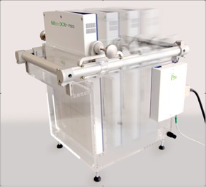
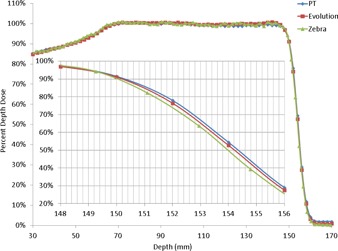
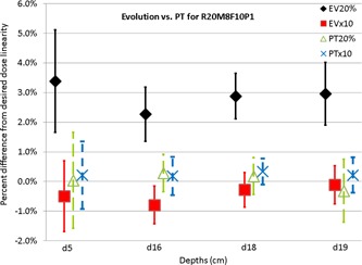
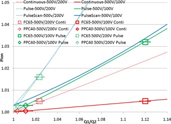
The need to accurately and efficiently verify both output and dose profiles creates significant challenges in quality assurance of pencil beam scanning (PBS) proton delivery. A system for PBS QA has been developed that combines a new two-dimensional ionization chamber array in a waterproof housing that is scanned in a water phantom. The MatriXX PT has the same detector array arrangement as the standard MatriXX(Evolution) but utilizes a smaller 2 mm plate spacing instead of 5mm. Because the bias voltage of the MatriXX PT and Evolution cannot be changed, PPC40 and FC65-G ionization chambers were used to assess recombination effects. The PPC40 is a parallel plate chamber with an electrode spacing of 2mm, while the FC65-G is a Farmer chamber FC65-G with an electrode spacing of 2.8 mm. Three bias voltages (500, 200, and 100 V) were used for both detectors to determine which radiation type (continuous, pulse or pulse-scanned beam) could closely estimate Pion from the ratios of charges collected. In comparison with the MatriXX(Evolution), a significant improvement in measurement of absolute dose with the MatriXX PT was observed. While dose uncertainty of the MatriXX(Evolution) can be up to 4%, it is < 1% for the MatriXX PT. Therefore the MatriXX(Evolution) should not be used for QA of PBS for conditions in which ion recombination is not negligible. Farmer chambers should be used with caution for measuring the absolute dose of PBS beams, as the uncertainty of Pion can be > 1%; chambers with an electrode spacing of 2 mm or smaller are recommended.
Figures
Similar articles
-
Design of a QA method to characterize submillimeter-sized PBS beam properties using a 2D ionization chamber array.Phys Med Biol. 2018 May 15;63(10):105007. doi: 10.1088/1361-6560/aabd89. Phys Med Biol. 2018. PMID: 29644984
-
Use of a two-dimensional ionization chamber array for proton therapy beam quality assurance.Med Phys. 2008 Sep;35(9):3889-94. doi: 10.1118/1.2963990. Med Phys. 2008. PMID: 18841839
-
Characterization and use of a 2D-array of ion chambers for brachytherapy dosimetric quality assurance.Med Dosim. 2012 Autumn;37(3):250-6. doi: 10.1016/j.meddos.2011.09.004. Epub 2011 Dec 19. Med Dosim. 2012. PMID: 22189031
-
Quality assurance of carbon ion and proton beams: A feasibility study for using the 2D MatriXX detector.Phys Med. 2016 Jun;32(6):831-7. doi: 10.1016/j.ejmp.2016.05.058. Epub 2016 May 28. Phys Med. 2016. PMID: 27246359
-
Calibration of a proton beam energy monitor.Med Phys. 2007 Jun;34(6):1952-66. doi: 10.1118/1.2717382. Med Phys. 2007. PMID: 17654898
Cited by
-
A Modular System for Treating Moving Anatomical Targets With Scanned Ion Beams at Multiple Facilities: Pre-Clinical Testing for Quality and Safety of Beam Delivery.Front Oncol. 2021 Mar 19;11:620388. doi: 10.3389/fonc.2021.620388. eCollection 2021. Front Oncol. 2021. PMID: 33816251 Free PMC article.
-
NRG Oncology Liver Proton SBRT and Hypofractionated Radiation Therapy: Current Treatment Technical Assessment and Practice Patterns.Cancers (Basel). 2025 Jul 17;17(14):2369. doi: 10.3390/cancers17142369. Cancers (Basel). 2025. PMID: 40723252 Free PMC article.
-
Nuclear halo measurements for accurate prediction of field size factor in a Varian ProBeam proton PBS system.J Appl Clin Med Phys. 2020 Jan;21(1):197-204. doi: 10.1002/acm2.12783. Epub 2019 Dec 2. J Appl Clin Med Phys. 2020. PMID: 31793202 Free PMC article.
-
Using field size factors to characterize the in-air fluence of a proton machine with a range shifter.Radiat Oncol. 2017 Mar 14;12(1):52. doi: 10.1186/s13014-017-0783-2. Radiat Oncol. 2017. PMID: 28288673 Free PMC article.
-
A multi-layer strip ionization chamber (MLSIC) device for proton pencil beam scan quality assurance.Phys Med Biol. 2022 Aug 23;67(17):10.1088/1361-6560/ac8593. doi: 10.1088/1361-6560/ac8593. Phys Med Biol. 2022. PMID: 35905730 Free PMC article.
References
-
- Arjomandy B, Sahoo N, Ding X, Gillin M. Use of a two‐dimensional ionization chamber array for proton therapy beam quality assurance. Med Phys. 2008;35(9):3889–95. - PubMed
-
- Arjomandy B, Sahoo N, Ciangaru G, Zhu R, Song X, Gillin M. Verification of patient‐specific dose distributions in proton therapy using a commercial two‐dimensional ion chamber array. Med Phys. 2010;37(11):5831–38. - PubMed
-
- Lomax A, Böhringer T, Bolsi A, et al. Treatment planning and verification of proton therapy using spot scanning: initial experiences. Med Phys. 2004;31(11):3150–57. - PubMed
-
- Pedroni E, Scheib S, Bohringer T, et al. Experimental characterization and physical modelling of the dose distribution of scanned proton pencil beams. Phys Med Biol. 2005;50(3):541–61. - PubMed
Publication types
MeSH terms
Substances
LinkOut - more resources
Full Text Sources
Other Literature Sources
