

Classification and clinicoradiologic features of primary progressive aphasia (PPA) and apraxia of speech
- PMID: 26103600
- PMCID: PMC4522343
- DOI: 10.1016/j.cortex.2015.05.013
Classification and clinicoradiologic features of primary progressive aphasia (PPA) and apraxia of speech
Abstract
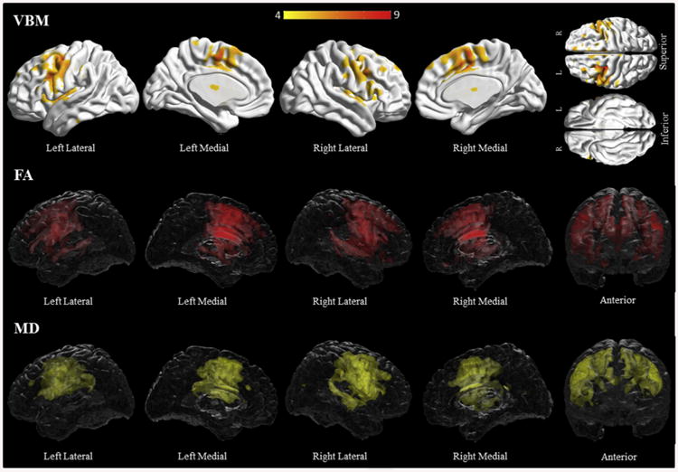
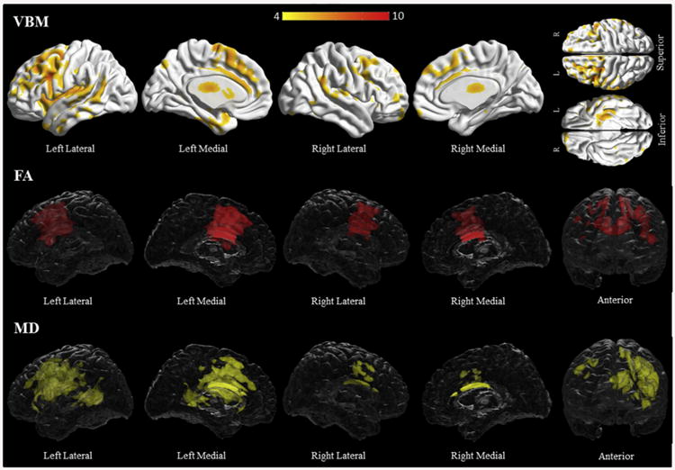
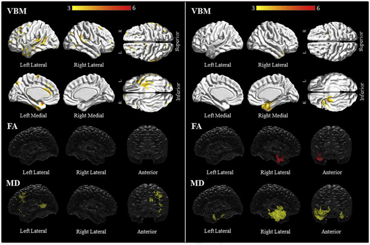
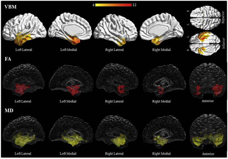
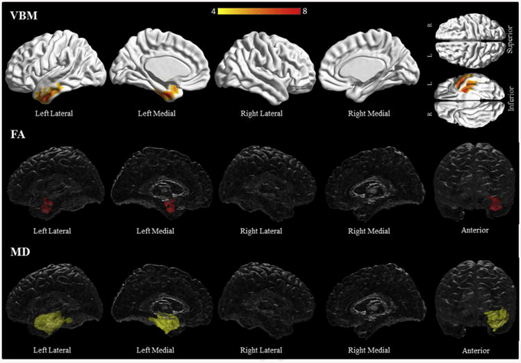
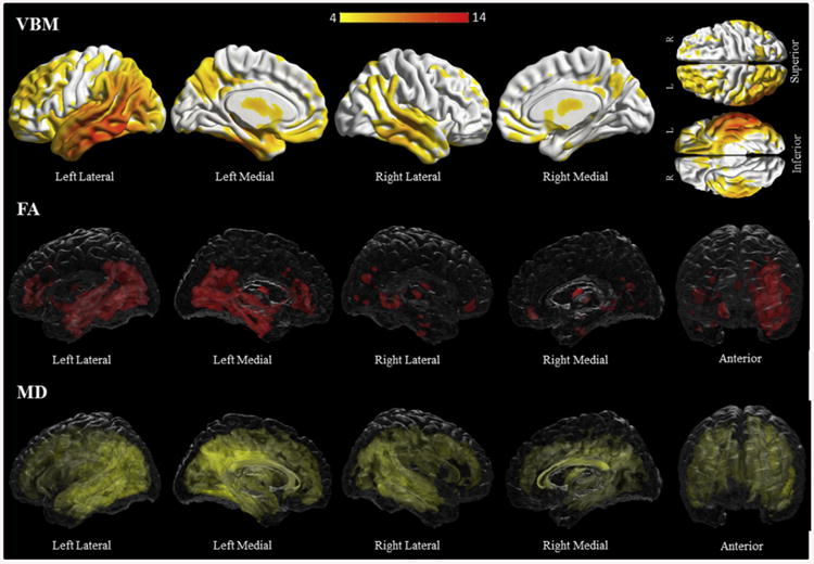
The consensus criteria for the diagnosis and classification of primary progressive aphasia (PPA) have served as an important tool in studying this group of disorders. However, a large proportion of patients remain unclassifiable whilst others simultaneously meet criteria for multiple subtypes. We prospectively evaluated a large cohort of patients with degenerative aphasia and/or apraxia of speech using multidisciplinary clinical assessments and multimodal imaging. Blinded diagnoses were made using operational definitions with important differences compared to the consensus criteria. Of the 130 included patients, 40 were diagnosed with progressive apraxia of speech (PAOS), 12 with progressive agrammatic aphasia, 9 with semantic dementia, 52 with logopenic progressive aphasia, and 4 with progressive fluent aphasia, while 13 were unclassified. The PAOS and progressive fluent aphasia groups were least impaired. Performance on repetition and sentence comprehension was especially poor in the logopenic group. The semantic and progressive fluent aphasia groups had prominent anomia, but only semantic subjects had loss of word meaning and object knowledge. Distinct patterns of grey matter loss and white matter changes were found in all groups compared to controls. PAOS subjects had bilateral frontal grey matter loss, including the premotor and supplementary motor areas, and bilateral frontal white matter involvement. The agrammatic group had more widespread, predominantly left sided grey matter loss and white matter abnormalities. Semantic subjects had bitemporal grey matter loss and white matter changes, including the uncinate and inferior occipitofrontal fasciculi, whereas progressive fluent subjects only had left sided temporal involvement. Logopenic subjects had diffuse and bilateral grey matter loss and diffusion tensor abnormalities, maximal in the posterior temporal region. A diagnosis of logopenic aphasia was strongly associated with being amyloid positive (46/52 positive). Our findings support consideration of an alternative way of identifying and categorizing subtypes of degenerative speech and language disorders.
Keywords: Amyloid PET imaging; Diffusion tensor imaging; Primary progressive aphasia; Progressive apraxia of speech; Volumetric based morphometry.
Copyright © 2015 Elsevier Ltd. All rights reserved.
Figures
References
-
- Adlam AL, Patterson K, Rogers TT, Nestor PJ, Salmond CH, Acosta-Cabronero J, et al. Semantic dementia and fluent primary progressive aphasia: two sides of the same coin? Brain. 2006;129:3066–3080. - PubMed
-
- Albert MS, DeKosky ST, Dickson D, Dubois B, Feldman HH, Fox NC, et al. The diagnosis of mild cognitive impairment due to Alzheimer's disease: recommendations from the National Institute on Aging-Alzheimer's Association workgroups on diagnostic guidelines for Alzheimer's disease. Alzheimer's & Dementia. 2011;7:270–279. - PMC - PubMed
-
- Alladi S, Xuereb J, Bak T, Nestor P, Knibb J, Patterson K, et al. Focal cortical presentations of Alzheimer's disease. Brain. 2007;130:2636–2645. - PubMed
Publication types
MeSH terms
Grants and funding
LinkOut - more resources
Full Text Sources
Other Literature Sources
