. 2015 Jun 24:6:7506.
doi: 10.1038/ncomms8506.

Exosomes released by keratinocytes modulate melanocyte pigmentation

Affiliations

Exosomes released by keratinocytes modulate melanocyte pigmentation

Alessandra Lo Cicero et al. Nat Commun. .

Abstract

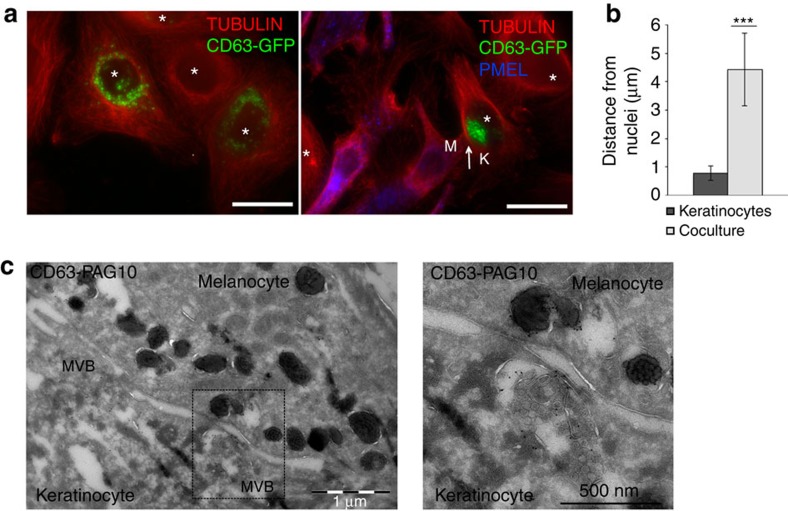
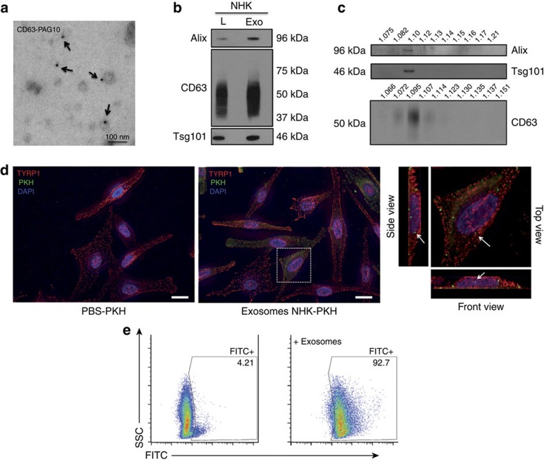
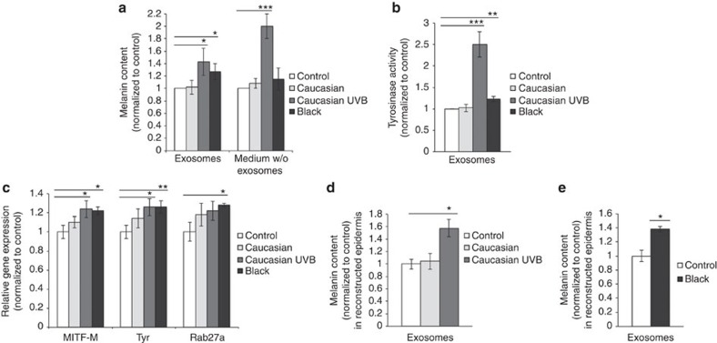
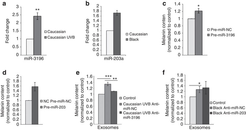
Cells secrete extracellular vesicles (EVs), exosomes and microvesicles, which transfer proteins, lipids and RNAs to regulate recipient cell functions. Skin pigmentation relies on a tight dialogue between keratinocytes and melanocytes in the epidermis. Here we report that exosomes secreted by keratinocytes enhance melanin synthesis by increasing both the expression and activity of melanosomal proteins. Furthermore, we show that the function of keratinocyte-derived exosomes is phototype-dependent and is modulated by ultraviolet B. In sum, this study uncovers an important physiological function for exosomes in human pigmentation and opens new avenues in our understanding of how pigmentation is regulated by intercellular communication in both healthy and diseased states.


Figures

Figure 1
Figure 1. MVB polarization in cell culture and reconstructed epidermis.
(a) CD63-GFP-transduced NHKs (green) in mono- or co-culture (ratio 1:1, melanocytes incubated with the same number of keratinocytes) with melanocytes were stained for tubulin (red) and PMEL (melanocyte-specific protein; blue) and were analysed using IFM (scale bar, 10 μm). Asterisks show the nuclei of keratinocytes and the arrow shows the site of contact between melanocyte and keratinocyte. (b) The distance of CD63-positive compartments from the centre of the corresponding nucleus was quantified in CD63-GFP-transduced NHK in mono- or co-culture with melanocytes (n=11; ***P<0.01, t-test). (c) EM analysis on ultrathin cryosections of Caucasian-reconstructed epidermis immunogold-labelled for endogenous CD63 (PAG 10 nm; scale bar, 1 μm). On the right, an inset corresponding to the magnified area of the back-boxed region depicts an MVB apposed to the keratinocyte plasma membrane in close association with melanocyte.
Figure 2
Figure 2. EV characterization and uptake.
(a) EM analysis of exosomes from NHK immunogold-labelled for endogenous CD63 (PAG 10 nm, arrows). (b) WB analysis of exosomes (Exo; 10 μg) and total cell lysate (L; 20 μg) from NHK or fractions recovered after OptiPrep gradient (c) using anti-Alix, -CD63 and -Tsg101 antibodies. (d) Analysis by IFM of the interaction of PKH67-labelled (green) exosomes from NHK with melanocytes labelled for TYRP1 (red) and DAPI (blue; scale bar, 10 μm). (e) FACS analysis of melanocytes incubated for 1 h at 37 °C with FITC-labelled exosomes from NHK. Melanocytes incubated with the last wash of the FITC-labelling were used as a control.
Figure 3
Figure 3. Exosomal effects on the regulation of pigmentation.
(a) Analysis of melanin content (optical density at 492 nm) in Caucasian melanocytes incubated for 96 h with NHK exosomes (corresponding to 15 μg of protein) resuspended in PBS (left) and medium depleted of NHK exosomes (right; Caucasian, Caucasian irradiated with ultraviolet B and Black; ratio 1:1, melanocytes incubated with exosomes or medium isolated from the same number of keratinocytes). Melanocytes incubated without exosomes were used as a control. (b,c) Tyrosinase activity (b) or relative gene expression of Mitf-M, Tyrosinase and Rab27a (c) were measured in Caucasian melanocytes incubated for 96 h with exosomes from NHK (Caucasian, Caucasian irradiated with ultraviolet B and Black; ratio 1:1, 15 μg of exosomes). Melanocytes incubated without exosomes were used as a control. Intracellular melanin content analysis of light phototype-reconstructed epidermis incubated with exosomes from Caucasian, ultraviolet B-irradiated Caucasian (d) or Black (e) NHK. Reconstructed epidermis cultured only with medium and PBS were used as controls. Experiments in d,e were performed with different epidermis of the same phototype using different melanocyte donors. The data are from three independent experiments. Values are mean±s.d. (*P<0.05; **P<0.02; ***P<0.01, t-test, n=3).
Figure 4
Figure 4. miRNA and pigmentation.
(a) Fold change of miR-3196 in exosomes from Black NHK and normalized to exosomes from Caucasian NHK. (b) Fold change of miR-203-a in exosomes from Caucasian-irradiated NHK and normalized to exosomes from Caucasian NHK. (cf) Analysis of intracellular melanin content in Caucasian melanocytes transfected with pre-miR-3196, (c) pre-miR-203 (d) or incubated for 96 h with exosomes from ultraviolet B-irradiated Caucasian NHK transfected with anti-miR-3196 (e) or with exosomes from Black NHK transfected with anti-miR-203. (f) Values are mean±s.d. (*P<0.05; **P<0.02; ***P<0.01, t-test, n=3).

Similar articles

Cited by

References

    1. Yamaguchi Y., Brenner M. & Hearing V. J. The regulation of skin pigmentation. J. Biol. Chem. 282, 27557–27561 (2007). - PubMed
    1. Yamaguchi Y. et al. Human skin responses to UV radiation: pigment in the upper epidermis protects against DNA damage in the lower epidermis and facilitates apoptosis. FASEB J. 20, 1486–1488 (2006). - PubMed
    1. Passeron T. et al. SOX9 is a key player in ultraviolet B-induced melanocyte differentiation and pigmentation. Proc. Natl Acad. Sci. USA 104, 13984–13989 (2007). - PMC - PubMed
    1. Cardinali G. et al. Melanosome transfer promoted by keratinocyte growth factor in light and dark skin-derived keratinocytes. J. Invest. Dermatol. 128, 558–567 (2008). - PubMed
    1. Raposo G. & Marks M. S. Melanosomes--dark organelles enlighten endosomal membrane transport. Nat. Rev. Mol. Cell Biol. 8, 786–797 (2007). - PMC - PubMed

Publication types

MeSH terms