. 2015 Dec;72(23):4653-69.
doi: 10.1007/s00018-015-1955-6. Epub 2015 Jun 24.

FAT1 cadherin acts upstream of Hippo signalling through TAZ to regulate neuronal differentiation

Affiliations

FAT1 cadherin acts upstream of Hippo signalling through TAZ to regulate neuronal differentiation

Abdulrzag F Ahmed et al. Cell Mol Life Sci. 2015 Dec.

Abstract

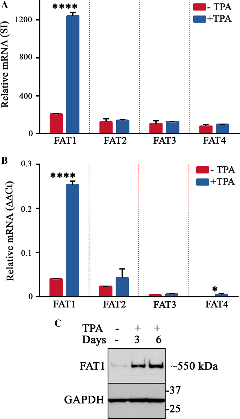
The Hippo pathway is emerging as a critical nexus that balances self-renewal of progenitors against differentiation; however, upstream elements in vertebrate Hippo signalling are poorly understood. High expression of Fat1 cadherin within the developing neuroepithelium and the manifestation of severe neurological phenotypes in Fat1-knockout mice suggest roles in neurogenesis. Using the SH-SY5Y model of neuronal differentiation and employing gene silencing techniques, we show that FAT1 acts to control neurite outgrowth, also driving cells towards terminal differentiation via inhibitory effects on proliferation. FAT1 actions were shown to be mediated through Hippo signalling where it activated core Hippo kinase components and antagonised functions of the Hippo effector TAZ. Suppression of FAT1 promoted the nucleocytoplasmic shuttling of TAZ leading to enhanced transcription of the Hippo target gene CTGF together with accompanying increases in nuclear levels of Smad3. Silencing of TAZ reversed the effects of FAT1 depletion thus connecting inactivation of TAZ-TGFbeta signalling with Hippo signalling mediated through FAT1. These findings establish FAT1 as a new upstream Hippo element regulating early stages of differentiation in neuronal cells.

Keywords: Cadherin; Differentiation; FAT1 cadherin; Hippo pathway; Neurite outgrowth; Neuronal differentiation; SMAD transcription factor; TAZ; TGFβ signalling.


Figures

Fig. 1
Fig. 1
FAT1 expression is strongly induced by differentiation of SH-SY5Y cells. a Expression microarray analysis of the relative mRNA levels of FAT family cadherins comparing untreated cells with those treated with TPA for 2 days. The mRNA levels of each FAT cadherin are expressed as the mean signal intensity (SI) ± SD of three biological replicates (****p ≤ 0.0001, t test). b Analysis of the relative mRNA levels of FAT family cadherins in SH-SY5Y cells left untreated for 6 days or treated with 80 nM TPA for 6 days using qPCR. The mRNA levels of each FAT cadherin are expressed as the mean ΔΔCt value ± SD from 3 replicates (see “Materials and methods”). Similar results were obtained in two experiments (*p ≤ 0.05; ****p ≤ 0.0001, t test). c Western blot analysis of FAT1 protein levels in cell lysates of SH-SY5Y cells untreated for 6 days or treated with 80 nM TPA for 3 and 6 days, respectively. Blots were probed with using affinity purified polyclonal antibodies raised against the cytoplasmic tail of FAT1 with similar results obtained in five experiments
Fig. 2
Fig. 2
Suppression of FAT1 induction during differentiation of SH-SY5Y cells decreases neurite outgrowth. a Western blotting analyses against cell lysates prepared from untreated cells or those treated with 80 nM TPA for 3 and 6 days, respectively. Comparison of cells transduced with control shRNA-miR (non-targeting sequence; NTS) or FAT1-targeting shRNA-miR demonstrates effective silencing of FAT1 protein expression. GAPDH was used as a loading control. b Representative phase contrast photomicrographs demonstrating phenotypic differences between neuritogenesis in control and FAT1-shRNA knockdown cells observed after 6 days of treatment with TPA. Bar represents 50 μm. c Quantitative comparison of the frequency and length of neurites in 6 day differentiated SH-SY5Y cell populations bearing NTS and FAT1-shRNA (n = 562 and 683 cells analysed, respectively). Neurites were defined as cellular projections as long or wide as the soma with absolute neurite lengths normalised against cell body measurements for each cell. Neurites were divided into six length categories ranging from 1 to 1.25 through to >5 cell body lengths as shown. d The percentage of cells from C recorded as having one or more neurites in either NTS or FAT1-shRNA populations (****p ≤ 0.0001, 2-sided Fisher’s exact test). e Box-Whisker plot showing neurite lengths for NTS and FAT1-shRNA populations. The box limits indicate the 25th and 75th percentiles with the internal line showing median values. Whiskers extend 1.5 times the interquartile range from the 25th and 75th percentiles (**p ≤ 0.01, Mann–Whitney U test). Plots were prepared using the BoxPlotR software package
Fig. 3
Fig. 3
Suppression of FAT1 expression following TPA-treatment affects cell density-dependent proliferation and survival. Cell growth rates were compared for NTS or FAT1-shRNA SH-SY5Y cells treated with 80 nM TPA. Cells were seeded at (a) low and (b) high densities (3750 and 15,000 cells/well, respectively) and total cell number estimated as fluorescence units determined through detection of co-transduced tGFP. Measurements conducted over consecutive days were normalised to day 1 with values representing relative fluorescence units (RFU) ± SD of six biological replicates. Similar results were obtained in three experiments (*p ≤ 0.05; ***p ≤ 0.001; ****p ≤ 0.0001, t test). c Comparative Western blotting analyses of FAT1 and cell cycle regulatory proteins (cyclin D1 and CDK2). Blots were performed on SH-SY5Y cell lysates prepared from NTS and two independent populations of FAT1-shRNA cells (refer Figure S2 and Table S2). GAPDH was used as a loading control
Fig. 4
Fig. 4
Expression of the Hippo pathway effectors YAP and TAZ during SH-SY5Y differentiation. a SH-SY5Y cells were treated with 80 nM TPA as described for Fig. 1c and cell lysate samples analysed for YAP and TAZ expression by Western blotting using a dual specificity polyclonal antibody recognising both YAP and TAZ. Anti- GAPDH was used as a loading control. b Transduced SH-SY5Y cells (NTS-control and FAT1 shRNA) populations were subjected to 80 nM TPA treatment or cultured without TPA as indicated. Cell lysate samples were subjected to Western blotting against YAP/TAZ. Thereafter, the membrane was reprobed with anti-GAPDH as a loading control
Fig. 5
Fig. 5
FAT1 acts through the core Hippo kinase cassette to inhibit nucleocytoplasmic shuttling of TAZ with regulatory effects on the levels and localisation of Smad3. a Subcellular fractionations enriched for nuclear or cytoplasmic proteins were prepared from NTS or FAT1-shRNA SH-SY5Y cells after 3 days of TPA treatment. Western blotting analyses were then performed using antibodies directed against TAZ, Smad2/3, p-Smad1/5/8 and Smad4. Lamin A/C and GAPDH were used as nuclear and cytoplasmic markers, respectively. b qPCR analysis of the Hippo pathway target gene, CTGF, was conducted on NTS or FAT1-shRNA SH-SY5Y cells after 3 days of TPA treatment. Prior to the assay, cells were pre-treated with either negative control (NC) or TAZ-targeted siRNA duplexes. c Western blotting analyses were performed against SH-SY5Y cell lysates using antibodies directed against the Hippo kinase cassette components (Mst1, Sav1 and p-MOB1) together with GAPDH as a loading control. d Western blotting analyses were performed against cell lysates prepared from NTS or FAT1-shRNA SH-SY5Y cells after 3 days of TPA treatment. Samples were blotted against Smad2/3 together with GAPDH as a loading control
Fig. 6
Fig. 6
The growth promoting effect and inhibition of neuritogenesis observed after FAT1 depletion are regulated through TAZ. a Control or FAT1-shRNA SH-SY5Y cells pre-treated with negative control (NC) or TAZ-targeted siRNA duplexes were treated with TPA for a period of 3 days. Western blotting analyses of cell lysates were performed against TAZ, Smad2/3 and the cell cycle regulatory proteins cyclin D1, CDK2 and CDK4. GAPDH was used as a loading control. b SH-SY5Y cell populations bearing NTS and FAT1-shRNA were pre-treated with control (NC) or TAZ-targeting siRNA duplexes prior to 6d of treatment with TPA. Thereafter neurite length was determined from >600 cells. Neurite lengths were normalised to cell body measurements for each cell and sub-divided into six length categories (1.000–1.25, 1.26–1.5, 1.51–2.00, 2.01–3.00, 3.01–5.00, >5.00) and plotted as histograms. c The percentage of cells recorded as having one or more neurites in indicated populations from B (a p ≤ 0.0001, b p ≤ 0.0001, c p ≤ 0.0001, Chi squared test). d Box-Whisker plot depicting neurite lengths from populations analysed in b. The box limits indicate the 25th and 75th percentiles with the internal line showing median values. Whiskers extend 1.5 times the interquartile range from the 25th and 75th percentiles [a p ≤ 0.0001, b p ≤ 0.01, c p ≤ 0.0001, d p ≤ 0.0001, e p ≤ 0.01, Kruskal–Wallis ANOVA by ranks test followed by post hoc analysis corrected for multiple comparisons (two-tailed)]. Plots were prepared using the BoxPlotR software package
Fig. 7
Fig. 7
Model depicting how FAT1 engages with the Hippo pathway to effect control of neuronal differentiation through antagonising TGFβ signalling. FAT1 is induced in cells triggered to differentiate where it promotes dual effects. First, FAT1 activates the core Hippo kinase cassette that serves to suppress the nuclear shuttling of TAZ. Second, FAT1 serves to both initiate and extend neurites (left panel). Conversely in the absence of FAT1 (or inhibiting its induction during differentiation using shRNA), Hippo kinases are not activated, thereby releasing constraints on TAZ. This serves to promote higher levels of Smad2/3 and both TAZ and Smad2/3 translocate to nucleus where they form transcriptionally active complexes involving TEADs and Smads. Activating gene targets of TEADs and Smad/TGFβ signalling invokes a transcriptional programme known to promote proliferation and self-renewal (right panel)
Fig. 8
Fig. 8
Expression analyses of Fat cadherins and TAZ in alternate models of neuronal differentiation. a Relative mRNA expression of Fat family cadherins (left) and TAZ (right) in murine embryonic brain tissue collected at the E9.5, E11.5 and E13.5 stages of development (GSE8091 dataset; [35]). The data represent the mean ± SD of 4-6 biological replicates per developmental stage. Each Fat cadherin was represented by one probe on the array while the results of two independent probes are shown for TAZ. b Gene expression analyses of FAT family cadherins (left) and TAZ (right) conducted on human iPSCs, neurospheres and neurons (GSE25542; [36]). The data represent the mean ± SD of 12 biological replicates per group. Probes for FAT3 were absent on the array. c Gene expression analyses of FAT family cadherins (left) and TAZ (right) conducted on human ESCs, neural rosettes and mature neurons (GSE40593; [37]). The data represent the mean ± SD of 3 biological replicates per differentiation stage. Probes for FAT3 were absent on the array while results for two independent probes are shown for TAZ. Statistical analyses were conducted by ANOVA followed by Tukeys Post HOC Test (ns not significant, a p < 0.05, b p < 0.01, c p < 0. 001, d p < 0. 0001)

References

    1. Sadeqzadeh E, de Bock CE, Thorne RF. Sleeping giants: emerging roles for the fat cadherins in health and disease. Med Res Rev. 2014;34(1):190–221. doi: 10.1002/med.21286. - DOI - PubMed
    1. Dunne J, Hanby AM, Poulsom R, Jones TA, Sheer D, Chin WG, Da SM, Zhao Q, Beverley PC, Owen MJ. Molecular cloning and tissue expression of FAT, the human homologue of the Drosophila fat gene that is located on chromosome 4q34-q35 and encodes a putative adhesion molecule. Genomics. 1995;30(2):207–223. doi: 10.1006/geno.1995.9884. - DOI - PubMed
    1. Matsui S, Utani A, Takahashi K, Mukoyama Y, Miyachi Y, Matsuyoshi N. Human Fat2 is localized at immature adherens junctions in epidermal keratinocytes. J Dermatol Sci. 2007;48(3):233–236. doi: 10.1016/j.jdermsci.2007.07.010. - DOI - PubMed
    1. Ciani L, Patel A, Allen ND, Ffrench-Constant C. Mice lacking the giant protocadherin mFAT1 exhibit renal slit junction abnormalities and a partially penetrant cyclopia and anophthalmia phenotype. Mol Cell Biol. 2003;23(10):3575–3582. doi: 10.1128/MCB.23.10.3575-3582.2003. - DOI - PMC - PubMed
    1. Saburi S, Hester I, Goodrich L, McNeill H. Functional interactions between Fat family cadherins in tissue morphogenesis and planar polarity. Development. 2012;139(10):1806–1820. doi: 10.1242/dev.077461. - DOI - PMC - PubMed

MeSH terms

Substances

LinkOut - more resources