Review
. 2015 Sep;24(9):1360-9.
doi: 10.1002/pro.2734. Epub 2015 Jul 7.

Structure-function correlations in tyrosinases

Affiliations
Review

Structure-function correlations in tyrosinases

Margarita Kanteev et al. Protein Sci. 2015 Sep.

Abstract

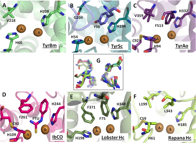
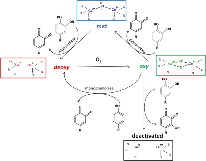
Tyrosinases are metalloenzymes belonging to the type-3 copper protein family which contain two copper ions in the active site. They are found in various prokaryotes as well as in plants, fungi, arthropods, and mammals and are responsible for pigmentation, wound healing, radiation protection, and primary immune response. Tyrosinases perform two sequential enzymatic reactions: hydroxylation of monophenols and oxidation of diphenols to form quinones which polymerize spontaneously to melanin. Two other members of this family are catechol oxidases, which are prevalent mainly in plants and perform only the second oxidation step, and hemocyanins, which lack enzymatic activity and are oxygen carriers. In the last decade, several structures of plant and bacterial tyrosinases were determined, some with substrates or inhibitors, highlighting features and residues which are important for copper uptake and catalysis. This review summarizes the updated information on structure-function correlations in tyrosinases along with comparison to other type-3 copper proteins.

Keywords: X-ray structure; catechol oxidase; copper; hemocyanin; type-3 copper proteins; tyrosinase.


Figures

Figure 1
Figure 1. The active site of type-3 copper enzymes
The active sites of type-3 copper proteins are shown in cartoon, active site residues are shown in sticks, and copper atoms are presented as brown spheres. (A) TyrBm active site and its blocker residue Val218 is shown in lime green. (B) The active site of TyrSc and its blocker residue Gly204 is shown in deep teal while the placeholder Tyr98 from the caddie protein is in blue. (C) The active site of TyrAo including Cys92 and His94 (thioether bond), a blocker residue Val359, and a placeholder Phe513 is presented in purple. (D) The active site of IbCO containing a thioehter bond between Cys92 and His109, a placeholder Phe261, and an inhibitor PTU is shown in pink. (E) Lobster Hc active site is presented in green with Phe371 and Phe75 as the blocker residue and a placeholder, respectively. (F) Rapana Hc active site is shown in lemon-green, containing a thioehter bond between Cys59 and His61, while Leu199 and Leu343 are the blocker residue and a placeholder, respectively. (G) Superposition of active site histidine residues of all type-3 copper proteins mentioned above. All figures were generated using PyMOL (http://www.pymol.org). Full names, PDB accession codes, and references are listed in Table I.
Figure 2
Figure 2. Oxidation states of tyrosinase
Four discrete states of the active site of tyrosinase are presented based on Ramsden and Riley (2014).5
Figure 3
Figure 3. Copper incorporation pathway
A) Copper transport from the caddie protein (blue color) to TyrSc (deep teal). B) The active site and second shell residues of TyrBm participating in copper transport. The active site and the second shell residues of TyrBm participating in copper transport. (C) The conserved motif of Cys residues participating in copper transport in TyrAo. (D) Two positions of CuB in abPPO4. Full names, PDB codes and references are listed in Table I.
Figure 4
Figure 4. Second shell residues of TyrBm
(A) A conserved methionine in a distance of 3.9Å from the second histidine residue coordinating CuB is important for stabilizing this His residue and enabling tyrosinase activity. (B) A structurally conserved water molecule participating in substrate deprotonation is ligated by conserved Glu196 and Asn205 residues. See Table I for respective residues in other type-3 copper proteins.
Figure 5
Figure 5. Similar orientation of tyrosine and l-Dopa in the active site of TyrBm
Superposition of the TyrBm structure with tyrosine (cyan, PDB accession code 4P6R) and the structure with l-Dopa (orange, PDB accession code 4P6S), showing similar orientation of monophenols and diphenols substrates in the active site of tyrosinases. The carboxyl side chains of the substrates form hydrogen bonds with Arg209. Zinc ions, replacing Cu ions in the crystal structure, are presented as grey spheres.

References

    1. Claus H, Decker H. Bacterial tyrosinases. Syst Appl Microbiol. 2006;29:3–14. - PubMed
    1. Faccio G, Kruus K, Saloheimo M, Thöny-Meyer L. Bacterial tyrosinases and their applications. Process Biochem. 2012;47:1749–1760.
    1. Fairhead M, Thony-Meyer L. Bacterial tyrosinases: old enzymes with new relevance to biotechnology. New Biotechnol. 2012;29:183–191. - PubMed
    1. Sanchez-Ferrer A, Rodriguez-Lopez JN, Garcia-Canovas F, Garcia-Carmona F. Tyrosinase: a comprehensive review of its mechanism. Biochim Biophys Acta. 1995;22:1–11. - PubMed
    1. Ramsden CA, Riley PA. Tyrosinase: the four oxidation states of the active site and their relevance to enzymatic activation, oxidation and inactivation. Biorg Med Chem. 2014;22:2388–2395. - PubMed

Publication types

Substances