Different tenogenic differentiation capacities of different mesenchymal stem cells in the presence of BMP-12
- PMID: 26104414
- PMCID: PMC4479325
- DOI: 10.1186/s12967-015-0560-7
Different tenogenic differentiation capacities of different mesenchymal stem cells in the presence of BMP-12
Abstract
Background: Mesenchymal stem cells (MSCs) are regarded as a promising cell-based therapeutic tool for tendon repair. This study aimed to compare the different tenogenic differentiation capacities of the three types of MSCs in the presence of bone morphogenic protein 12 (BMP-12).
Methods: MSCs were isolated from rat bone marrow (BM), inguinal adipose tissue (AD), and synovium (SM) from the knee joint. MSCs were characterized by morphology, proliferation, trilineage differentiation, and surface marker analysis. Tenogenic differentiation potential was initially assessed using real-time polymerase chain reaction, Western blot, and enzyme-linked immunosorbent assay in vitro. Histological assessments were also performed after subcutaneous implantation of BMP-12 recombinant adenovirus-infected MSCs in nude mice in vivo.
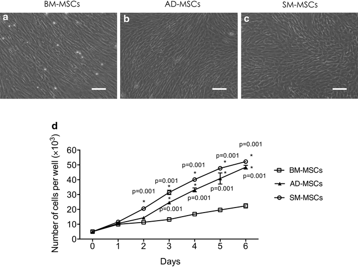
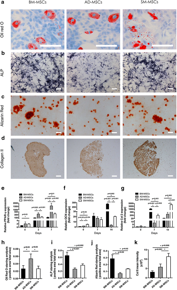
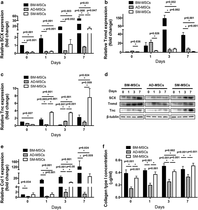
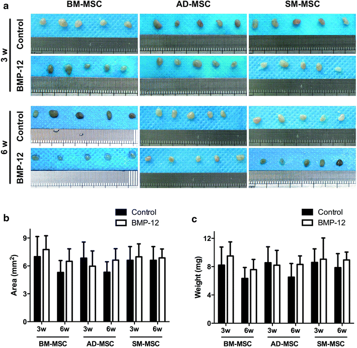
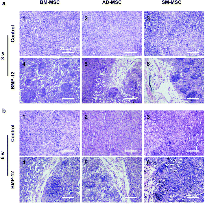
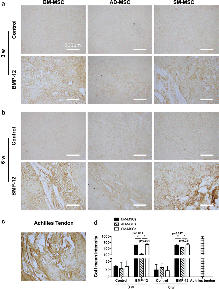
Results: The three types of MSCs exhibited similar fibroblast-like morphology and surface markers but different differentiation potentials toward adipogenic, osteogenic, and chondrogenic lineage fates. Bone marrow-derived MSCs (BM-MSCs) showed the most superior in vitro tenogenic differentiation capacity, followed by synovial membrane-derived MSCs (SM-MSCs) and then adipose-derived MSCs (AD-MSCs). After implantation, all three types of MSC masses infected with BMP-12 recombinant adenovirus emerged in the form of fiber-like matrix, especially in 6-week specimens, compared with the control MSCs in vivo. BM-MSCs and SM-MSCs revealed more intense staining for collagen type I (Col I) compared with AD-MSCs. Differences were not observed between BM-MSCs and SM-MSCs. However, SM-MSCs demonstrated higher proliferation capacity than BM-MSCs.
Conclusion: BM-MSCs exhibited the most superior tenogenic differentiation capacity, followed by SM-MSCs. By contrast, AD-MSCs demonstrated the inferior capacity among the three types of MSCs in the presence of BMP-12 both in vivo and in vitro.
Figures

Similar articles
-
Comparative study of the biological characteristics of mesenchymal stem cells from bone marrow and peripheral blood of rats.Tissue Eng Part A. 2012 Sep;18(17-18):1793-803. doi: 10.1089/ten.TEA.2011.0530. Epub 2012 Jul 30. Tissue Eng Part A. 2012. PMID: 22721583
-
Comparative analysis of human mesenchymal stem cells from bone marrow and adipose tissue under xeno-free conditions for cell therapy.Stem Cell Res Ther. 2015 Apr 13;6(1):55. doi: 10.1186/s13287-015-0066-5. Stem Cell Res Ther. 2015. PMID: 25884704 Free PMC article.
-
Mesenchymal stem cells from human bone marrow or adipose tissue differently modulate mitogen-stimulated B-cell immunoglobulin production in vitro.Cell Biol Int. 2008 Apr;32(4):384-93. doi: 10.1016/j.cellbi.2007.12.007. Epub 2008 Jan 9. Cell Biol Int. 2008. PMID: 18262807
-
Human bone marrow and adipose tissue mesenchymal stem cells: a user's guide.Stem Cells Dev. 2010 Oct;19(10):1449-70. doi: 10.1089/scd.2010.0140. Stem Cells Dev. 2010. PMID: 20486777 Review.
-
Same or not the same? Comparison of adipose tissue-derived versus bone marrow-derived mesenchymal stem and stromal cells.Stem Cells Dev. 2012 Sep 20;21(14):2724-52. doi: 10.1089/scd.2011.0722. Epub 2012 May 9. Stem Cells Dev. 2012. PMID: 22468918 Review.
Cited by
-
Biochemical Stimulus-Based Strategies for Meniscus Tissue Engineering and Regeneration.Biomed Res Int. 2018 Jan 17;2018:8472309. doi: 10.1155/2018/8472309. eCollection 2018. Biomed Res Int. 2018. PMID: 29581987 Free PMC article. Review.
-
The Lack of a Representative Tendinopathy Model Hampers Fundamental Mesenchymal Stem Cell Research.Front Cell Dev Biol. 2021 May 3;9:651164. doi: 10.3389/fcell.2021.651164. eCollection 2021. Front Cell Dev Biol. 2021. PMID: 34012963 Free PMC article. Review.
-
In vivo ligamentogenesis in embroidered poly(lactic-co-ε-caprolactone) / polylactic acid scaffolds functionalized by fluorination and hexamethylene diisocyanate cross-linked collagen foams.Histochem Cell Biol. 2023 Mar;159(3):275-292. doi: 10.1007/s00418-022-02156-3. Epub 2022 Oct 29. Histochem Cell Biol. 2023. PMID: 36309635 Free PMC article.
-
Current Progress in Tendon and Ligament Tissue Engineering.Tissue Eng Regen Med. 2019 Jun 26;16(6):549-571. doi: 10.1007/s13770-019-00196-w. eCollection 2019 Dec. Tissue Eng Regen Med. 2019. PMID: 31824819 Free PMC article. Review.
-
Growth factors in the treatment of Achilles tendon injury.Front Bioeng Biotechnol. 2023 Sep 14;11:1250533. doi: 10.3389/fbioe.2023.1250533. eCollection 2023. Front Bioeng Biotechnol. 2023. PMID: 37781529 Free PMC article. Review.
References
-
- Sharma P, Maffulli N. Biology of tendon injury: healing, modeling and remodeling. J Musculoskelet Neuronal Interact. 2006;6:181–190. - PubMed
-
- Hankemeier S, Keus M, Zeichen J, Jagodzinski M, Barkhausen T, Bosch U, et al. Modulation of proliferation and differentiation of human bone marrow stromal cells by fibroblast growth factor 2: potential implications for tissue engineering of tendons and ligaments. Tissue Eng. 2005;11:41–49. doi: 10.1089/ten.2005.11.41. - DOI - PubMed
Publication types
MeSH terms
Substances
LinkOut - more resources
Full Text Sources
Other Literature Sources

