Review
. 2015 Feb;3(1):MDNA3-0050-2014.
doi: 10.1128/microbiolspec.MDNA3-0050-2014.

Mobile Bacterial Group II Introns at the Crux of Eukaryotic Evolution

Affiliations
Review

Mobile Bacterial Group II Introns at the Crux of Eukaryotic Evolution

Alan M Lambowitz et al. Microbiol Spectr. 2015 Feb.

Abstract

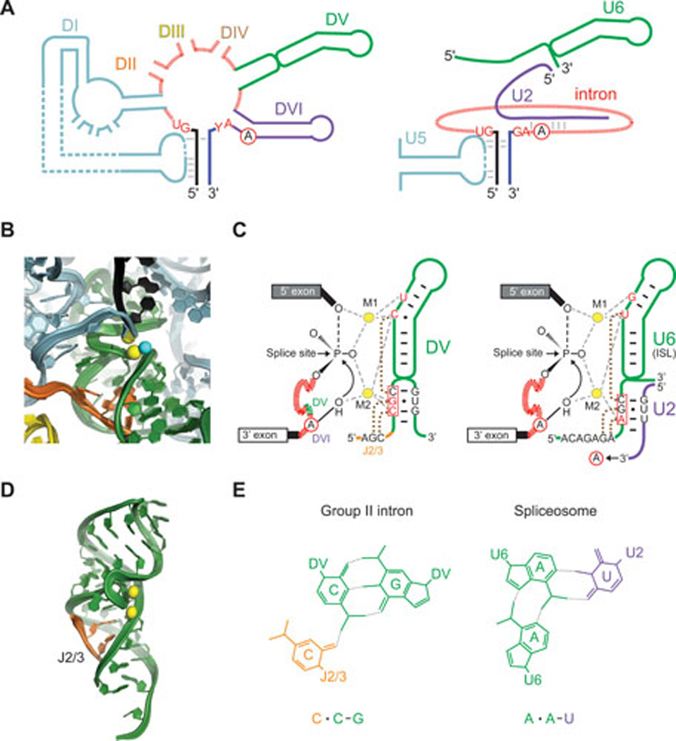
This review focuses on recent developments in our understanding of group II intron function, the relationships of these introns to retrotransposons and spliceosomes, and how their common features have informed thinking about bacterial group II introns as key elements in eukaryotic evolution. Reverse transcriptase-mediated and host factor-aided intron retrohoming pathways are considered along with retrotransposition mechanisms to novel sites in bacteria, where group II introns are thought to have originated. DNA target recognition and movement by target-primed reverse transcription infer an evolutionary relationship among group II introns, non-LTR retrotransposons, such as LINE elements, and telomerase. Additionally, group II introns are almost certainly the progenitors of spliceosomal introns. Their profound similarities include splicing chemistry extending to RNA catalysis, reaction stereochemistry, and the position of two divalent metals that perform catalysis at the RNA active site. There are also sequence and structural similarities between group II introns and the spliceosome's small nuclear RNAs (snRNAs) and between a highly conserved core spliceosomal protein Prp8 and a group II intron-like reverse transcriptase. It has been proposed that group II introns entered eukaryotes during bacterial endosymbiosis or bacterial-archaeal fusion, proliferated within the nuclear genome, necessitating evolution of the nuclear envelope, and fragmented giving rise to spliceosomal introns. Thus, these bacterial self-splicing mobile elements have fundamentally impacted the composition of extant eukaryotic genomes, including the human genome, most of which is derived from close relatives of mobile group II introns.


Figures

Figure 1
Figure 1. Group II intron RNA splicing mechanism and structure
A. Splicing and reverse splicing. Step 1. The 2’-OH of the branch-point adenosine acts as nucleophile to attack the 5' splice site. Step 2. The 3'-OH of the upstream exon is the nucleophile that attacks the 3' splice site to generate ligated exons and an excised intron lariat. Both reactions are reversible. B. Group II intron secondary structure. The L. lactis Ll.LtrB group IIA intron is shown, with the six domains DI-DVI. Exons are represented by thicker lines with the EBS-IBS pairings shown by dashed black lines. The IEP ORF is looped out of DIV (not drawn to scale). C. Group II intron crystal structure. The representation is of DI-DV of the O. iheyensis group IIC intron (PDB:4E8K) bound to ligated exons, before the spliced exon reopening reaction, provided by Marco Marcia and Anna Pyle (13). Colors are coded to the domain labels in panel B, although these are different introns that belong to different structural subgroups. The 5' exon is black, and the 3' exon is dark blue. D. Base-pairing interactions of group IIA, IIB and IIC introns with flanking exons. Group IIA and IIB recognize 5' exons by similar IBS1-EBS1 and IBS2-EBS2 interactions, but use different interactions to recognize 3' exons (δ-δ' in IIA introns and EBS3/IBS3 in IIB introns (213). Group IIC ribozymes are only ∼400 nt long, considerably smaller than IIA and IIB introns, and they are located downstream of inverted repeats, such as transcription terminators, which contribute to exon recognition along with short EBS1/IBS1 and EBS3/IBS3 interactions similar to those of IIB introns (214, 215). Panel D is adapted from reference (4), with permission of the publisher (copyright Cold Spring Harbor Laboratory Press).
Figure 2
Figure 2. Group II intron and related RTs
A. Schematics of RTs. Two group II intron RTs, L. lactis Ll.LtrB (denoted LtrA protein; GenBank: AAB06503) and Sinorhizobium meliloti RmInt (NCBI Reference Sequence: NP_438012) are compared with two non-LTR-retrotransposon RTs, Bombyx mori R2Bm (GenBank: AAB59214) and human LINE-1 (UniProtKB/Swiss-Prot: O00370) RTs; yeast telomerase RT (GenBank: AAB64520); and retrovirus HIV-1 RT (PDB:2HMI). Conserved sequence blocks in the RT domain are numbered, and the sequence motif containing two of the conserved aspartic acid residues at the RT active site is shown below for each protein. B. Three-dimensional model of the Ll.LtrB RT. Regions identified by unigenic evolution analysis as being required for binding the high-affinity binding site DIVa and catalytic core regions of group II intron RNAs are highlighted in red and dark blue in the left and right panels, respectively, with pink in the left panel indicating a region that may enhance DIVa binding by stabilizing the structure of neighboring regions (46). The model was constructed by threading the amino acid sequence of the Ll.LtrB RT onto a HIV-1 RT crystal structure, with one subunit (denoted α; gray) modeled based on the catalytic p66 subunit of HIV-1 RT and the other subunit (denoted β; cyan) modeled based on the p51 subunit of HIV-1 RT (17). The N-terminal 36 amino acid residues of the Ll.LtrB RT could not be modeled based on the HIV-1 RT and are represented as spheres. Abbreviations: APE, apurinic endonuclease domain; CTS, conserved carboxy-terminal segment found to bind RNA non-specifically in LINE-1 RTs (216); Cys, cysteine-rich sequence conserved in LINE-1 RTs; DB, DNA-binding domain in R2Bm RT; REL, restriction endonuclease-like domain; TEN, telomerase N-terminal domain; TRBD, telomerase RNA-binding domain including motifs CP and T, which contact telomerase RNA (139, 217).
Figure 3
Figure 3. Representative retrohoming and retrotransposition pathways
A and B. Retrohoming into cognate sites is represented by the two pathways on the left. C and D. Retrotransposition to ectopic sites is represented by the two pathways on the right. For all panels, the intron is red (DNA solid lines, RNA hatched lines); the intron RNP is represented by a grey rectangle with a red intron lariat and an IEP with RT and maturase (X) domains and either containing or lacking an En domain. Exons of the donor (encircled D) are white, and those of the recipient (encircled R) are either white (retrohoming) or grey (retrotransposition). Each pathway ends with a product (encircled P). Green exon fragment represents primer for reverse transcription. The retrohoming pathways (A and B) differ by the presence of the En domain, and whether dsDNA (A) or ssDNA, such as at a replication fork (B), is the target. Black dot in the recipient strand represents the intron-insertion site. In pathway A, the IEP contains an En domain and after reverse splicing into the top strand, cleavage of the bottom strand occurs 9 or 10 nt downstream (step 1'), as for the Ll.LtrB and yeast aI2 introns, respectively (54, 101). In pathway B, the IEP lacks an En domain and integrates into DNA at a replication fork (step 1) as demonstrated for RmInt1 intron (62) and En-deficient mutants of the Ll.LtrB intron (78). Reverse splicing into the leading strand is not shown (see (78)). In both pathways, cDNA synthesis proceeds with the intron as template, using the 3'-OH of either the cleaved bottom strand (A) or an Okazaki fragment (B) to prime reverse transcription (step 2). Intron degradation and second-strand cDNA synthesis (step 3) is followed by DNA repair to generate the retrohoming products for both pathways (step 4). Host factors that participate in the process are indicated on pathway A, as established for the Ll.LtrB intron in E. coli, with those that silence the pathway shown in red, and those that promote retrohoming indicated in green (Table 1) (72, 73). EPP, error-prone polymerase. Retrotransposition (C and D) occurs when the intron integrates into ectopic sites with reduced specificity. This occurs for the Ll.LtrB intron into the lagging strand template for DNA synthesis, as pathway B, where Okazaki fragments prime cDNA synthesis (C) (58). Stimulatory and repressive host factors are again represented in green and red, respectively (Table 1) (45, 74, 97). Alternatively primers can be provided by nicks introduced into dsDNA by relaxase, the product of the ltrB gene that hosts the intron (pathway D) (93). Steps 1–4 in pathway D are as for retrohoming.
Figure 4
Figure 4. DNA target site recognition by group IIA, IIB and IIC introns
Target site interactions are shown for retrohoming (100, 101, 103) and retrotransposition (59, 92, 93) of the Ll.LtrB group IIA intron, and for retrohoming of group IIB introns EcI5 (104) and RmInt1 (77), and group IIC intron B.h.I1-B (107). Intron RNA regions involved in EBS1-IBS1, EBS2-IBS2, δ-δ’ or EBS3-IBS3 base-pairing interactions with the DNA target site are shown in red. A representative ectopic site is shown for the Ll.LtrB retrotransposition pathway. Base-pairs in the 5' and 3' exons that are recognized by the IEP are highlighted in mauve and blue, respectively. CS, bottom-strand cleavage site; IS, intron-insertion site; RF, replication fork; RH, retrohoming; RTP, retrotransposition; SL, stem-loop. CS for EcI5 is not known.
Figure 5
Figure 5. Similar RNA active sites of group II introns and the spliceosome
A. Schematic of group II intron (left) and spliceosome (right). Group II intron elements are colored as in Figure 1B and C, with corresponding spliceosomal elements in the same color (D1 and U5 blue; DV and U6 green; DVI and U2, purple). Conserved residues at the splice sites are in red, and the branch-point adenosine (A) is circled. Elements are not drawn to scale. B. Crystal structure of O. iheyensis group II intron in the precatalytic state (from PDB file 4FFAQ). The 5' exon (black) is shown before 5' splice site hydrolysis. Color-coding is as in Figure 1B & C and Figure 5A, with the two catalytic Mg2+ shown as yellow spheres bound to DV (green) (13, 218). The putative water nucleophile is a cyan sphere. Images in panel B and D provided by Marco Marcia and Anna Pyle. C. Secondary structures and Mg2+ interactions in O. iheyensis intron DV and spliceosomal U6 snRNA. DV (left, shown also in panel B) corresponds to the internal stem loop (ISL) of U6 (right). M1 and M2 are catalytic Mg2+ ions coordinated by phosphate oxygens of the nucleotides shown in red. The circled A in the intron is the adenosine nucleophile with the 2’-OH corresponding to the water molecule in B, which attacks the 5' splice site (arrow). The introns are depicted in red hatch marks, with a small segment in green representing DV of the group II intron detailed to the right. U2 3' to the U2-U6 pairing extends to interact with the branch-point region of the intron (arrow to circled A). The three boxed nucleotides in each case comprise the conserved catalytic triad (163, 178, 183, 219). Dotted brown lines join residues involved in base triples, which are formed by two pairings between the catalytic triad and ether J2/3 or the 5'-ACAGAGA-3' box and the third pairing to the bulge in each structure (163, 180). D. Base triples in DV of the O. iheyensis intron. The J2/3 nucleotides (orange ribbon) form a triple helix with the major groove at the base of DV (from (10)). Two metal ions (yellow spheres) are bound near the twisted bulge loop. E. Example of base triples in DV and U2-U6. The color-coded base triples are shown for the lower-most base-pair in the catalytic triad with a nucleotide in J2/3 in the group II intron and in the 5'-ACAGAGA-3' box in U6 of the spliceosome, as diagramed in panel C.
Figure 6
Figure 6. Comparison of Prp8 to group II intron and related RTs
Schematic comparing S. cerevisiae Prp8 (PDB:4I43), with the Ll.LtrB group II intron RT (LtrA protein; GenBank:AAB06503), Arabidopis thaliana nMat1 and nMat2 proteins (NCBI Reference Sequence: NP_174294 and NP_177575, respectively), non-LTR-retrotranspson R2Bm RT (GenBank:AAB59214) and Neurospora crassa RVT RT (GenBank:CAE76174). The plant nMat1 and nMat2 proteins are previous examples of group II intron RTs that were subsumed into nuclear genomes and retained RNA splicing function, with the nMat1 proteins acquiring a novel conserved domain (green) in place of the En domain. The Prp8 configuration of the thumb, long linker, and REL domain is similar to that in the R2Bm RT. Although group II intron RTs and the R2Bm RT lack an RNase H domain, RNase H domains are known to be acquired sporadically in different non-LTR-retrotransposon lineages (135, 191). RVT RTs, another potential candidate for an ancestor of Prp8 (181), lack En and integrase domains and are further distinguished from group II intron and non-LTR-retrotransposon RTs by a large acidic insertion within RT-2a and conserved N- and C-terminal domains that are not found in other proteins (212). Conserved sequence blocks in the RT domain are numbered, and the sequence motif containing two of the conserved aspartic acid residues at the RT active site is shown below for each protein. The locations of conserved RT sequence blocks in Prp8 are from structure-based sequence alignments by Georg Mohr. Abbreviations: DB, DNA binding domain; REL, restriction endonuclease-like domain.

Similar articles

Cited by

References

    1. Lambowitz AM, Zimmerly S. Mobile group II introns. Annu Rev Genet. 2004;38:1–35. - PubMed
    1. Pyle AM, Lambowitz AM. The RNA World. 3rd ed. Cold Spring Harbor Laboratory Press; 2006. Group II introns: ribozymes that splice RNA and invade DNA; pp. 469–505.
    1. Toro N, Jiménez-Zurdo JI, García-Rodríguez FM. Bacterial group II introns: not just splicing. FEMS Microbiol Rev. 2007;31:342–358. - PubMed
    1. Lambowitz AM, Zimmerly S. Group II introns: mobile ribozymes that invade DNA. Cold Spring Harbor Perspectives in Biology. 2011;3:a003616. - PMC - PubMed
    1. Marcia M, Somarowthu S, Pyle AM. Now on display: a gallery of group II intron structures at different stages of catalysis. Mobile DNA. 2013;4:14. - PMC - PubMed

Publication types

LinkOut - more resources