. 2015 Jun 24:15:196.
doi: 10.1186/s12906-015-0700-x.

Effects of Ixeris dentata water extract and caffeic acid on allergic inflammation in vivo and in vitro

Affiliations

Effects of Ixeris dentata water extract and caffeic acid on allergic inflammation in vivo and in vitro

Yong-Deok Jeon et al. BMC Complement Altern Med. .

Abstract

Background: Ixeris dentata Nakai has been used for the treatment of mithridatism, calculous, indigestion, pneumonia, hepatitis, and tumors in Korea, China, and Japan. However, the effect of a water extract of Ixeris dentata (ID) and its molecular mechanism on allergic inflammation has not been elucidated. In this study, we attempted to evaluate the effects of ID and its major compound caffeic acid on allergic inflammation in vivo and in vitro.

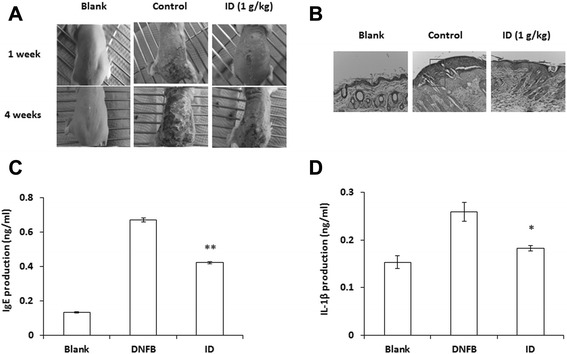
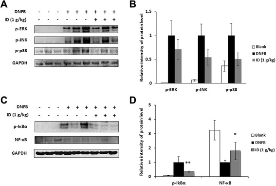
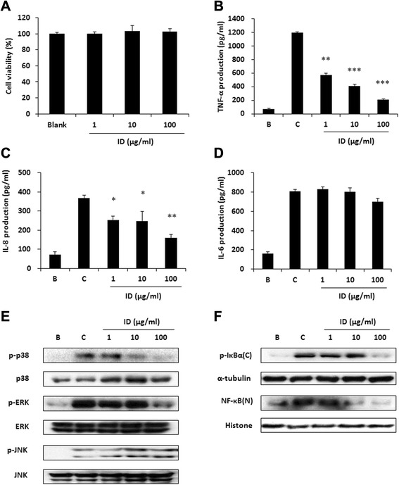
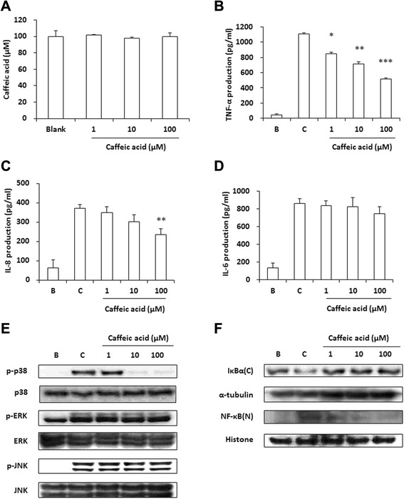
Methods: ID was applied to 2, 4-dinitrofluorobenzene (DNFB)-induced atopic dermatitis (AD)-like skin lesion mice and immune cell infiltration, cytokine production, and the activation of mitogen-activated protein kinases (MAPKs) were investigated. Moreover, the effect of ID on compound 48/80-induced anaphylactic shock was investigated in a mouse model. The human keratinocyte cell line (HaCaT cells) and human mast cells (HMC-1) were treated with ID or caffeic acid to investigate the effects on the production of chemokines and proinflammatory cytokines and on the activation of MAPKs.

Results: ID inhibited the serum levels of IgE and interleukin (IL)-1β in DNFB-induced AD-like skin lesion mouse models and suppressed anaphylactic shock in the mouse models. ID and caffeic acid inhibited the production of chemokines and adhesion molecules in HaCaT cells. In addition, ID reduced the release of tumor necrosis factor-α and IL-8 via the inhibition of MAPKs phosphorylation in HMC-1 cells.

Conclusions: These results suggest that ID is a potential therapeutic agent for allergic inflammatory diseases, including dermatitis.


Figures

Fig. 1
Fig. 1
Effects of ID on DNFB-induced dermatitis and serum IgE and IL-1β levels. BALB/c mice (n = 7) were sensitized with 100 μl of 0.15 % DNFB in an acetone-olive oil combination (3: 1) or a vehicle (acetone/olive oil = 3: 1) applied to the dorsal skin twice each week for a total period of six weeks. ID (1 g/kg) was orally administrated to DNFB-treated mice for six weeks. (a) Comparison of DNFB-induced dermatitis in BALB/c mice after oral administration of ID. (b) H&E stained tissue sections. Skin tissues were fixed with 10 % formaldehyde embedded in paraffin. (c and d) Blood samples were collected from the mice, and the levels of serum IgE (c) and IL-1β (d) in the indicated groups were measured with the ELISA method. Values represent the mean ± SD of independent experiments. . *p < 0.05, **p < 0.01
Fig. 2
Fig. 2
Effects of ID on activation of MAPKs and NF-κB in skin lesions. Protein was isolated from normal or DNFB-induced dermatitis dorsal skin. (a) Phosphorylation levels of ERK, JNK, and p38 was assayed by western blot analysis. (b) Relative levels of p-ERK, p-JNK, and p-p38 were calculated using an image analyzer. Values represent the mean ± SD of independent experiments. (c) The phosphorylation of IκBα and the protein expressions of NF-κB were assayed by western blot analysis. (d) The relative levels of p-IκBα and NF-κB were calculated using an image analyzer. Values represent the mean ± SD of independent experiments. *p < 0.05, **p < 0.01
Fig. 3
Fig. 3
Effects of ID and caffeic acid on the expression levels of inflammatory mediators in HaCaT cells. (a and b) HaCaT cells (2 × 105 cells/well) were treated with ID (0.01 - 1 mg/ml) or caffeic acid (1–100 μM) for 12 h and the degree of cell viability was analyzed by an MTT assay. (c and d) HaCaT cells (2 × 105 cells/well) were pre-treated with ID or caffeic acid for 30 min and were stimulated with TNF-α/IFN-γ for 18 h. The levels of cytokine were assayed by a RT-PCR analysis. (e and f) Relative levels of IL-8, TARC, MDC, ICAM-1, and MMP-9 were calculated using an image analyzer. Values represent the mean ± SD of three independent experiments. CA, caffeic acid. *p < 0.05, **p < 0.01
Fig. 4
Fig. 4
Effects of ID on the production of proinflammatory cytokines and the activation of MAPKs and NF-κB in HMC-1 cells. (a) HMC-1 cells (1 × 104 cells/well) were treated with ID (0.01 - 1 mg/ml) for 24 h and the cell viability was analyzed with MTT assays. (b-d) The production levels of TNF-α (B), IL-8 (c), and IL-6 (d) were measured using the ELISA method. HMC-1 cells (2 × 105 cells/well) were pre-treated with ID for 1 h and were stimulated with PMA (50 nM) + A23187 (1 μM) for 8 h. Values represent the mean ± SD of three independent experiments. *p < 0.05, **p < 0.01, ***p < 0.001. (e and f) Phosphorylation of p38, ERK and JNK (e) and the phosphorylation of IκBα and translocation of NF-κB (f) were assayed by western blot analysis. HMC-1 cells (1 × 106 cells/well) were incubated with various concentrations of ID and then stimulated with PMA (50 nM) + A23187 (1 μM) for 30 min (e) or 2 h (f). C, cytosol extracts; N, nuclear extracts
Fig. 5
Fig. 5
Effects of caffeic acid on the production of proinflammatory cytokines and the activation of MAPKs and NF-κB in HMC-1 cells. (a) HMC-1 cells (1 × 104 cells/well) were treated with caffeic acid (1–100 μM) for 24 h and the degree of cell viability was analyzed with MTT assays. (b-d) The production levels of TNF-α (b), IL-8 (c), and IL-6 (d) were measured using the ELISA method. HMC-1 cells (2 × 105 cells/well) were pre-treated with caffeic acid for 1 h and were stimulated with PMA (50 nM) + A23187 (1 μM) for 8 h. Values represent the mean ± SD of three independent experiments. *p < 0.05, **p < 0.01, ***p < 0.001. (e and f) Phosphorylation of p38, ERK and JNK (e) and the degradation of IκBα and translocation of NF-κB (f) were assayed by western blot analysis. HMC-1 cells (1 × 106 cells/well) were incubated with various concentrations of caffeic acid and then stimulated with PMA (50 nM) + A23187 (1 μM) for 30 min (e) or 2 h (f). C, cytosol extracts; N, nuclear extracts

References

    1. Leung DY, Bieber T. Atopic dermatitis. Lancet. 2003;361:151–160. doi: 10.1016/S0140-6736(03)12193-9. - DOI - PubMed
    1. Leung DY, Boguniewicz M, Howell MD, Nomura I, Hamid QA. New insights into atopic dermatitis. J Clin Invest. 2004;113:651–657. doi: 10.1172/JCI21060. - DOI - PMC - PubMed
    1. Bonness S, Bieber T. Molecular basis of atopic dermatitis. Curr Opin Allergy Clin Immunol. 2007;7:382–386. doi: 10.1097/ACI.0b013e3282a643c3. - DOI - PubMed
    1. Novak N. New insights into the mechanism and management of allergic diseases: atopic dermatitis. Allergy. 2009;64:265–275. doi: 10.1111/j.1398-9995.2008.01922.x. - DOI - PubMed
    1. Ahn EM, Bang MH, Song MC, Park MH, Kim HY, Kwon BM, Baek NI. Cytotoxic and ACAT-inhibitory sesquiterpene lactones from the root of Ixeris dentata forma albiflora. Arch Pharm Res. 2006;29:937–941. doi: 10.1007/BF02969274. - DOI - PubMed

Publication types

LinkOut - more resources