Review
. 2015 Apr;3(2):10.1128/microbiolspec.PLAS-0036-2014.
doi: 10.1128/microbiolspec.PLAS-0036-2014.

Topological Behavior of Plasmid DNA

Affiliations
Review

Topological Behavior of Plasmid DNA

N Patrick Higgins et al. Microbiol Spectr. 2015 Apr.

Abstract

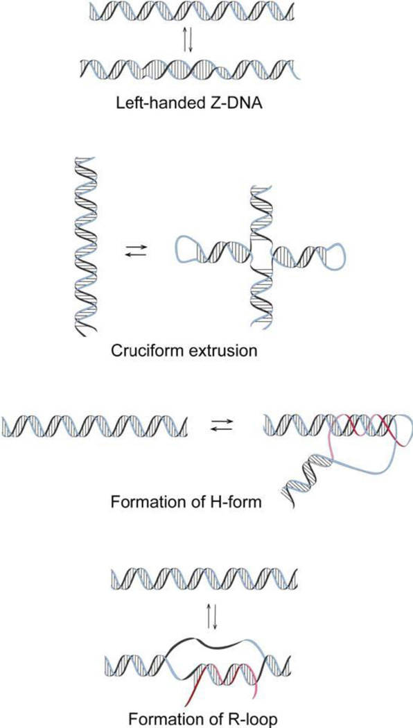
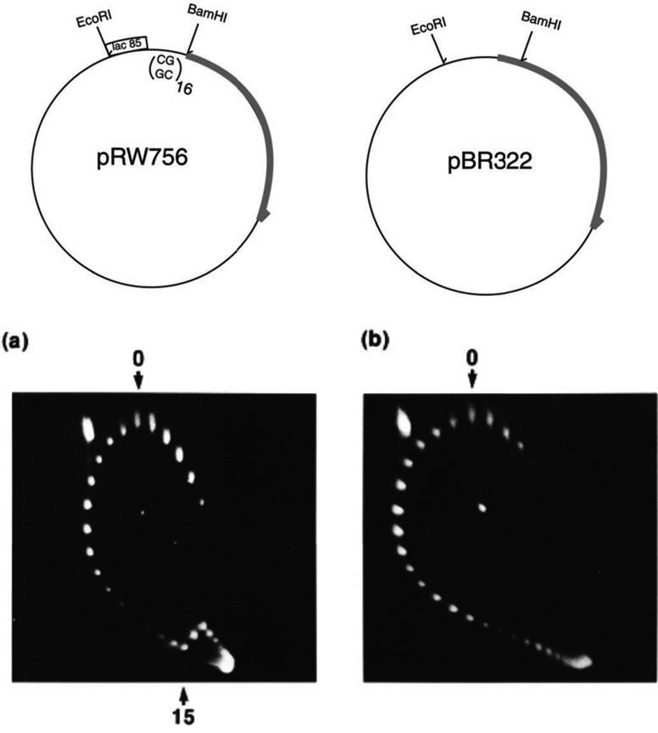
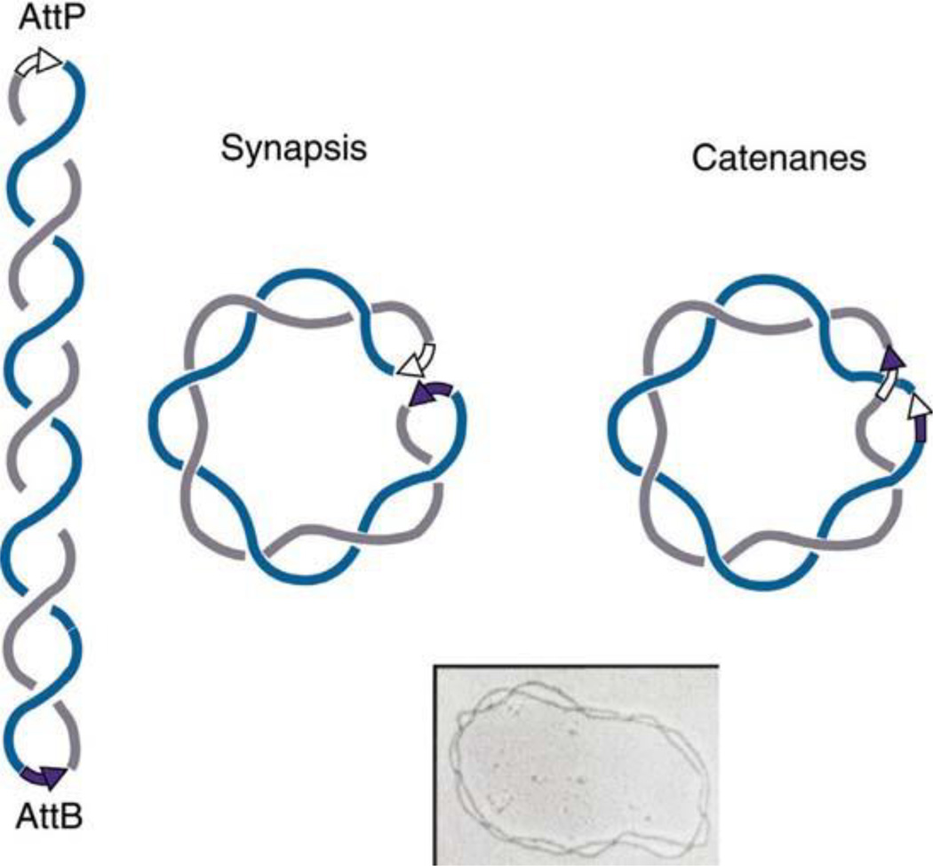
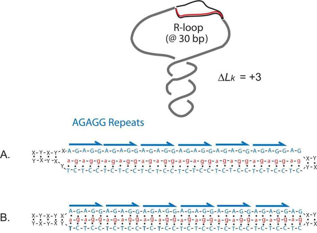
The discovery of the B-form structure of DNA by Watson and Crick led to an explosion of research on nucleic acids in the fields of biochemistry, biophysics, and genetics. Powerful techniques were developed to reveal a myriad of different structural conformations that change B-DNA as it is transcribed, replicated, and recombined and as sister chromosomes are moved into new daughter cell compartments during cell division. This article links the original discoveries of superhelical structure and molecular topology to non-B form DNA structure and contemporary biochemical and biophysical techniques. The emphasis is on the power of plasmids for studying DNA structure and function. The conditions that trigger the formation of alternative DNA structures such as left-handed Z-DNA, inter- and intra-molecular triplexes, triple-stranded DNA, and linked catenanes and hemicatenanes are explained. The DNA dynamics and topological issues are detailed for stalled replication forks and for torsional and structural changes on DNA in front of and behind a transcription complex and a replisome. The complex and interconnected roles of topoisomerases and abundant small nucleoid association proteins are explained. And methods are described for comparing in vivo and in vitro reactions to probe and understand the temporal pathways of DNA and chromosome chemistry that occur inside living cells.


Conflict of interest statement

Conflicts of interest: We declare no conflicts.

Figures

FIGURE 1
FIGURE 1
The simplest knots (a) and catenanes (b). DNA molecules are capable of adopting these and many more complex topological states. doi:10.1128/microbiolspec.PLAS-0036-2014.f1
FIGURE 2
FIGURE 2
Diagram of closed circular DNA. The linking number, Lk, of the complementary strands is 18. doi:10.1128/microbiolspec.PLAS-0036-2014.f2
FIGURE 3
FIGURE 3
Typical simulated conformations of supercoiled DNA 4.4 kb in length. The conformations correspond to a DNA superhelix density of (a) −0.030 and (b) −0.060. doi:10.1128/microbiolspec.PLAS-0036-2014.f3
FIGURE 4
FIGURE 4
Electrophoretic separation of topoisomers of pUC19 DNA. The mixture of topoisomers covering the range of ΔLk from 0 to −8 was electrophoresed from a single well in 1% agarose that was run from top to bottom. The topoisomer with ΔLk = 0 has the lowest mobility: it moves slightly slower than the open circular form (OC). The value of (−ΔLk) for each topoisomer is shown. doi:10.1128/microbiolspec.PLAS-0036-2014.f4
FIGURE 5
FIGURE 5
Separation of pUC19 DNA topoisomers by two-dimensional gel electrophoresis. Topoisomers 1 to 4 are positively supercoiled; the rest have negative supercoiling. After electrophoresis in the first direction, from top to bottom, the gel was saturated with ligand intercalating into the double helix. Upon electrophoresis from left to right in the second direction, the 2nd and 13th topoisomers turned out to migrate near the relaxed position in the second dimension. The spot in the top-left corner corresponds to the open circular form (OC); the spot near the middle of the gel corresponds to linear DNA (L). doi:10.1128/microbiolspec.PLAS-0036-2014.f5
FIGURE 6
FIGURE 6
High-resolution gel electrophoresis of knotted forms of plasmid DNA that was run from left to right. Knot types are described in Fig. 1 (see reference 21). Reproduced from the Journal of Molecular Biology with permission from Elsevier. doi:10.1128/microbiolspec.PLAS-0036-2014.f6
FIGURE 7
FIGURE 7
Alternative DNA structures that are stabilized by negative supercoiling. doi:10.1128/microbiolspec.PLAS-0036-2014.f7
FIGURE 8
FIGURE 8
Two-dimensional gel showing the transition from B- to left-handed Z-DNA in plasmid DNA. This research was originally published in Kang DS, Wells RD. 1985. B-Z DNA junctions contain few, if any, nonpaired bases at physiological superhelical densities. J Biol Chem 260:7783-7790. © the American Society for Biochemistry and Molecular Biology. doi:10.1128/microbiolspec.PLAS-0036-2014.f8
FIGURE 9
FIGURE 9
Conversion of interwound negative supercoils into catenanes linked by site-specific recombination. EM reprinted from Spergler SJ, Stasiak A, Cozzarelli NR. 1985. The stereo-structure of knots and catenanes produced by phage lambda integrative recombination: implications for mechanism and DNA structure. Cell 42:325-334 with permission from Elsevier. doi:10.1128/microbiolspec.PLAS-0036-2014.f9
FIGURE 10
FIGURE 10
Alternative RNA-DNA structures that contribute to constrained supercoiling in a plasmid containing a fragment of the chicken IgA immunoglobulin switch region during transcription. The R-loop structure shown in (A) results in a displaced strand of DNA that constrains a ΔLk of about +1 for every 10 bp of RNA/DNA hybrid. (B) shows the structure of an intermolecular triplex in which Hoogsteen base pairing occurs in the major groove of the DNA strand (see reference 159). doi:10.1128/microbiolspec.PLAS-0036-2014.f10
FIGURE 11
FIGURE 11
Proposed mechanism for stable RNA-DNA hybrids that can stimulate repeat instability. Transcription of DNA regions containing CG-rich trinucleotide repeats (red) favors formation of stable RNA-DNA hybrids. The displaced nontemplate DNA strand can adopt non-B DNA structures, such as CTG or CAG hairpins. The unpaired regions of the nontemplate strand are reactive to bisulfite modification. Reprinted from reference with permission from the National Academy of Sciences. doi:10.1128/microbiolspec.PLAS-0036-2014.f11
FIGURE 12
FIGURE 12
Replication intermediates identified in plasmid replication systems. (A) Replication initiated at a unique position leads to dual forms that move toward the terminus of replication. (B) Introduction of positive supercoils leads to replication fork reversal and formation of a four-way junction. (C) Negative supercoiling, which is generated by gyrase ahead of the fork, can be converted into precatenanes (D), which become catenanes (E) upon completion of DNA synthesis. (F, G) Topoisomerase activity in the replicated region can lead to complex knots. doi:10.1128/microbiolspec.PLAS-0036-2014.f12
FIGURE 13
FIGURE 13
Model of replication repair. Strand displacement and branch migration create an alternative replication template allowing replication to bypass a lesion (X). Reproduced from reference with permission from Elsevier. doi:10.1128/microbiolspec.PLAS-0036-2014.f13
FIGURE 14
FIGURE 14
Replication fork reversal in vivo (see Fig. 12). Reprinted from reference with permission from the American Association for the Advancement of Science. doi:10.1128/microbiolspec.PLAS-0036-2014.f14
FIGURE 15
FIGURE 15
Resolution of catenane (CATS) and precatenane links (RI) in plasmid DNA (see Fig. 12). Reprinted from reference with permission from Elsevier. doi:10.1128/microbiolspec.PLAS-0036-2014.f15
FIGURE 16
FIGURE 16
Schematic models for generating hemicatenanes during DNA replication. Three pathways to yield hemicatenane structures are shown. (Left) Lagging strand synthesis encounters a damage site, and the pairing of the lagging strand with the complementary leading strand can produce a pseudo-double Holliday structure. Dissolution of the pseudo-double Holliday structure leads to hemicatenanes and allows replication to bypass the damage site. (Center) Convergence of two replication forks at the final stage of replication can lead to either a single-strand catenane or hemicatenane conjoining two replicated duplexes. Both single-strand catenanes and hemicatenanes can be resolved by a type IA topoisomerase, allowing the segregation of the daughter chromosomes. (Right). Convergent branch migration of a double Holliday junction can generate a hemicatenane. Reproduced from reference with permission from the National Academy of Sciences. doi:10.1128/microbiolspec.PLAS-0036-2014.f16
FIGURE 17
FIGURE 17
Knotting of replication bubbles in vivo. Reprinted from reference with permission from Wiley. doi:10.1128/microbiolspec.PLAS-0036-2014.f17

References

    1. Vinograd J, Lebowitz J, Radloff R, Watson R, Laipis P. The twisted circular form of polyoma viral DNA. Proc Natl Acad Sci USA. 1965;53:1104–1111. - PMC - PubMed
    1. Bauer WR, Crick FHC, White JH. Supercoiled DNA. Sci Am. 1980;243:100–113. - PubMed
    1. Calugareanu G. Sur las classes d’isotopie des noeuds tridimensionnels et leurs invariants. Czech Math J. 1961;11:588–625.
    1. White JH. Self-linking and the Gauss integral in higher dimensions. Am J Math. 1969;91:693–728.
    1. Fuller FB. The writhing number of a space curve. Proc Natl Acad Sci USA. 1971;68:815–819. - PMC - PubMed

Publication types

LinkOut - more resources