Review
. 2015 Apr;3(2):MDNA3-0034-2014.
doi: 10.1128/microbiolspec.MDNA3-0034-2014.

Mechanisms of DNA Transposition

Affiliations
Review

Mechanisms of DNA Transposition

Alison B Hickman et al. Microbiol Spectr. 2015 Apr.

Abstract

DNA transposases use a limited repertoire of structurally and mechanistically distinct nuclease domains to catalyze the DNA strand breaking and rejoining reactions that comprise DNA transposition. Here, we review the mechanisms of the four known types of transposition reactions catalyzed by (1) RNase H-like transposases (also known as DD(E/D) enzymes); (2) HUH single-stranded DNA transposases; (3) serine transposases; and (4) tyrosine transposases. The large body of accumulated biochemical and structural data, particularly for the RNase H-like transposases, has revealed not only the distinguishing features of each transposon family, but also some emerging themes that appear conserved across all families. The more-recently characterized single-stranded DNA transposases provide insight into how an ancient HUH domain fold has been adapted for transposition to accomplish excision and then site-specific integration. The serine and tyrosine transposases are structurally and mechanistically related to their cousins, the serine and tyrosine site-specific recombinases, but have to date been less intensively studied. These types of enzymes are particularly intriguing as in the context of site-specific recombination they require strict homology between recombining sites, yet for transposition can catalyze the joining of transposon ends to form an excised circle and then integration into a genomic site with much relaxed sequence specificity.


Figures
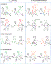
FIGURE 1
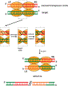
FIGURE 1
Basic chemical reactions catalyzed by DNA transposases. (A) An RNase H-like active site, based on structures of PFV intasomes (28, 94, 95). The green DNA represents the cleaved dinucleotide, and orange is the target strand. Spheres indicate bound metal ions. (B) HUH nuclease active site acting on single-stranded DNA (based on PDB ID 2X06 of ISDra2 TnpA). Shown is the reaction that occurs at the transposon Left End (LE). After cleavage, the DNA flanking the LE (black) remains in the active site; upon exchange of α-helices between the two active sites of the dimeric transposase, the cleaved LE moves to the other monomer where it is joined to the cleaved RE to form a circular excised transposon (not shown). At the same time, the flanking DNA from the RE of the transposon switches active sites (as shown here in black) and subsequent joining results in a sealed donor backbone. (C) DNA cleavage catalyzed by a serine recombinase. The active serine is surrounded by many Arg residues. Upon 180° rotation of one dimer within a tetramer, one strand rotates out of the active site (green) while another rotates in (orange). (D) DNA cleavage catalyzed by a tyrosine recombinase. Crucial residues within the active site include a conserved RHR triad (for details, see also (181)). doi:10.1128/microbiolspec.MDNA3-0034-2014.f1
FIGURE 2
FIGURE 2
Proposed pathway for transposon circle integration into target DNA catalyzed by a serine transposase. At the top, a tetrameric assembly is shown bringing together the abutted Left End (LE; orange) and Right End (RE; red) of an excised circular transposon with a target DNA (green). The reactions in the dashed box show how four cleavage reactions in which each active site serine becomes covalently attached to one strand of DNA, followed by a 180 degree rotation of the left-most dimer, leads to a reorganization of the strands. Resolution of the four covalent intermediates results in an integrated transposon. doi:10.1128/microbiolspec.MDNA3-0034-2014.f2
FIGURE 3
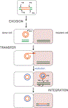
FIGURE 3
Pathway of conjugative transposition. Whether catalyzed by a serine or a tyrosine transposase, excision results in a circular intermediate in which the transposon ends are abutted. Only one of the strands of this intermediate is transferred to the recipient cell, and replication (new strands shown in blue) regenerates the double-stranded form in both cells. doi:10.1128/microbiolspec.MDNA3-0034-2014.f3
FIGURE 4
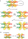
FIGURE 4
Proposed pathways of excision and integration by tyrosine transposases. (A) Transposon excision. (B) Transposon integration. doi:10.1128/microbiolspec.MDNA3-0034-2014.f4
FIGURE 5
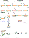
FIGURE 5
Transposition pathways for RNase H-like transposases. Arrows indicate sites of strand cleavage and the black dots indicate 3′-OH groups. Many pathways converge on essentially the same form of the excised transposon (highlighted with grey boxes). This linear intermediate is then integrated into target DNA as shown in (f). Target site duplications (TSDs) are generated when the cell repairs the gaps introduced by staggered strand transfer reactions. Adapted from (1). doi:10.1128/microbiolspec.MDNA3-0034-2014.f5
FIGURE 6
FIGURE 6
Transpososome structures for the RNase H-like DNA transposases. Cartoon representations of five transpososomes containing RNase H-like catalytic domains determined by X-ray crystallography. In all images, the catalytically active protomers acting on the two transposon ends are colored in orange and green, with the green catalytic domain acting on the DNA end shown in blue and the orange domain acting on the DNA end in red. Where target DNA is present, it is shown in grey. Inactive protomers (MuA, PFV IN and Hermes) are colored purple and magenta. The following PDB codes were used: (A) Tn5, 1MUH; (B) PFV IN, 4E7J; (C) Mos1, 3HOT; (D) MuA, 4FCY; (E) Hermes, 4D1Q. doi:10.1128/microbiolspec.MDNA3-0034-2014.f6
FIGURE 7
FIGURE 7
Transpososome of the HUH transposase TnpA of IS608, modelled as binding one Left End (LE; red) and one Right End (RE; blue). The PDB codes 2VJV and 2VJU were used. The inset shows the step of the reaction in the strand transfer and reset model for IS608 transposition (see He et al., this volume) to which the structure corresponds; note that the RE flank has not yet been observed crystallographically. doi:10.1128/microbiolspec.MDNA3-0034-2014.f7

References

    1. Curcio MJ, Derbyshire KM. 2003. The outs and ins of transposition: From Mu to kangaroo. Nature Rev Mol Cell Biol 4:865–877. - PubMed
    1. Montaño SP, Rice PA. 2011. Moving DNA around: DNA transposition and retroviral integration. Curr Opin Struct Biol 21:370–378. - PMC - PubMed
    1. Chandler M, de la Cruz F, Dyda F, Hickman AB, Moncalian G,Ton-Hoang B. 2013. Breaking and joining single-stranded DNA: the HUH endonuclease superfamily. Nature Rev Microbiol 11:525–538. - PMC - PubMed
    1. Yang W 2011. Nucleases: diversity of structure, function and mechanism. Quart Rev Biophys 44:1–93. - PMC - PubMed
    1. Dyda F, Hickman AB, Jenkins TM, Engelman A, Craigie R,Davies DR. 1994. Crystal structure of the catalytic domain of HIV-1 integrase: Similarity to other polynucleotidyl transferases. Science 266: 1981–1986. - PubMed

Publication types

LinkOut - more resources