Review
. 2016 Jun;15(3):369-91.
doi: 10.1007/s12311-015-0687-3.

Consensus Paper: Revisiting the Symptoms and Signs of Cerebellar Syndrome

Affiliations
Review

Consensus Paper: Revisiting the Symptoms and Signs of Cerebellar Syndrome

Florian Bodranghien et al. Cerebellum. 2016 Jun.

Abstract

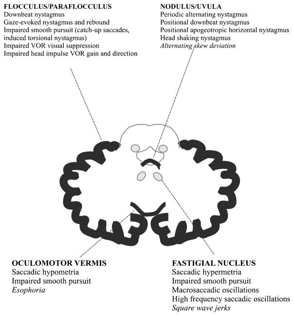
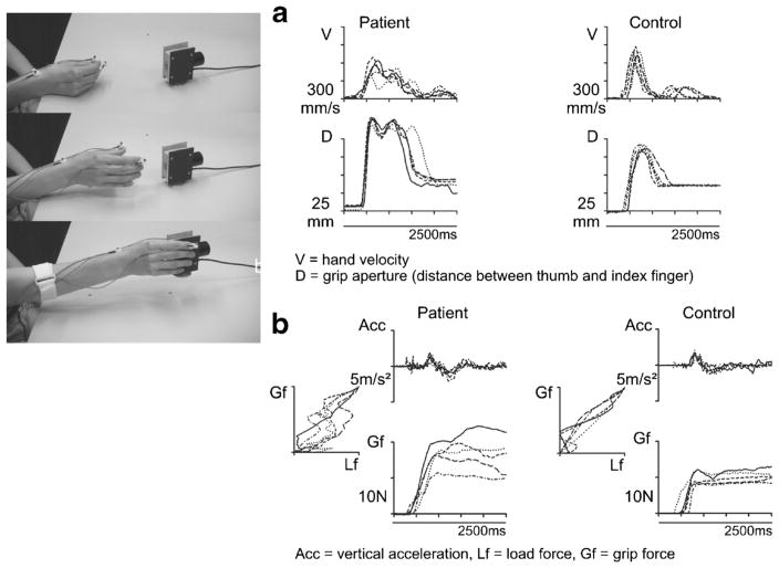
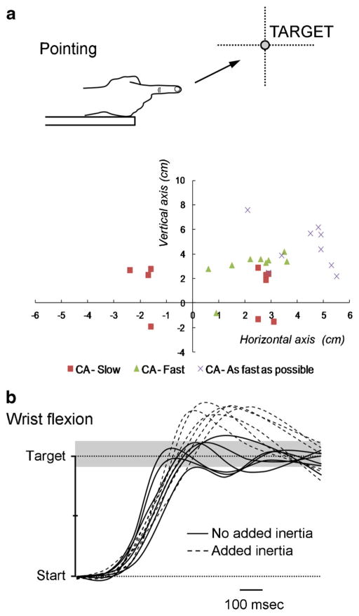
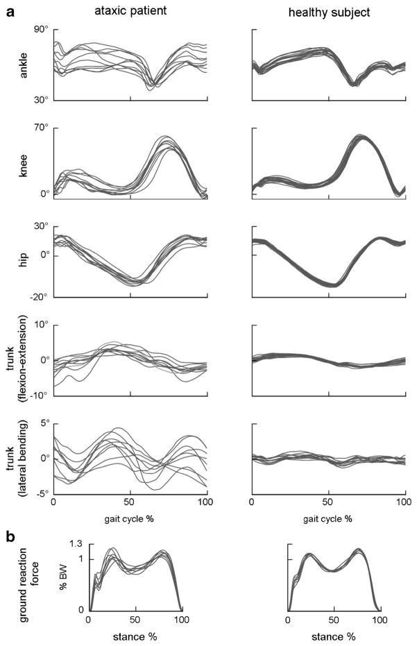
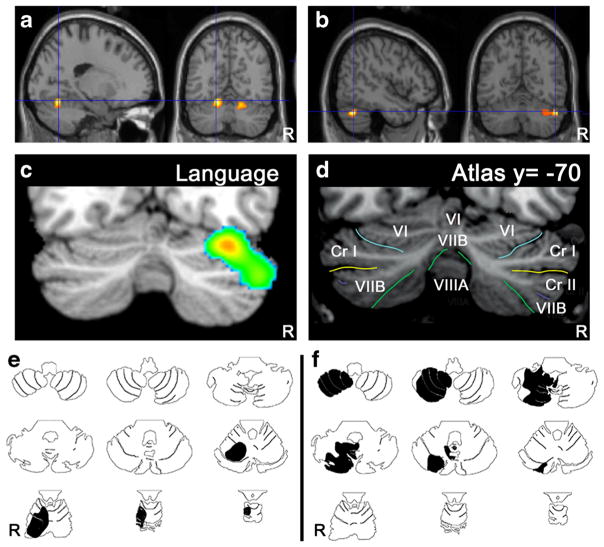
The cerebellum is involved in sensorimotor operations, cognitive tasks and affective processes. Here, we revisit the concept of the cerebellar syndrome in the light of recent advances in our understanding of cerebellar operations. The key symptoms and signs of cerebellar dysfunction, often grouped under the generic term of ataxia, are discussed. Vertigo, dizziness, and imbalance are associated with lesions of the vestibulo-cerebellar, vestibulo-spinal, or cerebellar ocular motor systems. The cerebellum plays a major role in the online to long-term control of eye movements (control of calibration, reduction of eye instability, maintenance of ocular alignment). Ocular instability, nystagmus, saccadic intrusions, impaired smooth pursuit, impaired vestibulo-ocular reflex (VOR), and ocular misalignment are at the core of oculomotor cerebellar deficits. As a motor speech disorder, ataxic dysarthria is highly suggestive of cerebellar pathology. Regarding motor control of limbs, hypotonia, a- or dysdiadochokinesia, dysmetria, grasping deficits and various tremor phenomenologies are observed in cerebellar disorders to varying degrees. There is clear evidence that the cerebellum participates in force perception and proprioceptive sense during active movements. Gait is staggering with a wide base, and tandem gait is very often impaired in cerebellar disorders. In terms of cognitive and affective operations, impairments are found in executive functions, visual-spatial processing, linguistic function, and affective regulation (Schmahmann's syndrome). Nonmotor linguistic deficits including disruption of articulatory and graphomotor planning, language dynamics, verbal fluency, phonological, and semantic word retrieval, expressive and receptive syntax, and various aspects of reading and writing may be impaired after cerebellar damage. The cerebellum is organized into (a) a primary sensorimotor region in the anterior lobe and adjacent part of lobule VI, (b) a second sensorimotor region in lobule VIII, and (c) cognitive and limbic regions located in the posterior lobe (lobule VI, lobule VIIA which includes crus I and crus II, and lobule VIIB). The limbic cerebellum is mainly represented in the posterior vermis. The cortico-ponto-cerebellar and cerebello-thalamo-cortical loops establish close functional connections between the cerebellum and the supratentorial motor, paralimbic and association cortices, and cerebellar symptoms are associated with a disruption of these loops.

Keywords: A- or Dysdiadochokinesia; Affect; Ataxia; Cerebellar syndrome; Cerebellum; Cognition; Dysarthria; Dysmetria; Eye movements; Functional topography; Hypotonia; Language; Loops; Speech; Tremor.


Conflict of interest statement

Conflicts Of Interest The authors declare no relevant conflict of interest.

Figures

Fig. 1
Fig. 1
Structural-clinical correlation of oculomotor deficits associated with cerebellar disorders. In italics, provisional localization. Adapted from Leigh and Zee [40]
Fig. 2
Fig. 2
Rapid alternate movements (RAMs) in cerebellar disorders. The figure is taken from Holmes [6]. RAMs are slowed on the left (L) side, ipsilaterally to a gunshot injury of the cerebellum, compared to the right (R) side. Increasing variability in movement frequency and amplitude is also seen, which becomes more prominent during the course of the movements due to fatigue (permission granted)
Fig. 3
Fig. 3
a Profiles of hand transport velocity and grasp formation during repetitive reaching to grasp movements performed by a subject with cerebellar degeneration and a healthy control subject. b Profiles of lifting acceleration and grip force during repetitive grasp and lift movements performed by a subject with cerebellar degeneration and a healthy control subject. Modified according to Brandauer et al. [86]
Fig. 4
Fig. 4
a Pointing movements of the right upper limb in a patient presenting with a stroke in the territory of the right superior cerebellar artery (CA). End movements (corresponding to the first zero-crossing value of the velocity curve) are represented. The target is located at a distance of 85 % of the upper limb length, at the height of the shoulder. Series of 10 movements performed at slow speed (CA-Slow red squares), at fast speed (CA-Fast green triangles) and as fast as possible (CA-As fast as possible X). Note that the initial dysmetria (motion at slow speed) is transformed into a genuine hypermetria (overshoot) as the velocity increases. b Fast goal-directed wrist movements in a patient with a cerebellar stroke which is clinically silent. Adding inertia (+300 g) triggers an overshoot of the target (dotted lines) located at 20° from the start position. Gray zone corresponds to the range of control values
Fig. 5
Fig. 5
Examples of joint and trunk orientation angles (a) and vertical ground reaction force (b) in one ataxic patient (left) and one age-, sex- and gait speed-matched control (right) during overground walking. Every trace refers to a single gait cycle. Stride-by-stride temporal variability of both kinematic and kinetic patterns is consistently larger in ataxic gait
Fig. 6
Fig. 6
Topographic arrangement in cerebellum of speech versus language representation. Functional MRI localizes articulation (a) to medial parts of lobule VI bilaterally, whereas verb generation (b) activates lateral regions of lobule VI and crus I on the right. In a meta-analysis of functional imaging studies [156] higher-level language tasks engage the right lateral posterior cerebellum, lobules VI and crus I (c) according to the lobule identification in (d; [184]). Case studies of cerebellar stroke patients reveal topography for articulation vs. higher-level language tasks. A patient with stroke in the territory of the right superior cerebellar artery (e, black shading) involving lobules I–VI was dysarthric; whereas a patient with stroke in the territory of the right posterior inferior cerebellar artery (f, black shading) involving lobules VII–IX was not dysarthric but performed poorly on the Boston Naming Test [185] (from Stoodley and Schmahmann in ref. [11])

References

    1. Koziol LF, Budding D, Andreasen N, D’Arrigo S, Bulgheroni S, Imamizu H, et al. Consensus paper: the cerebellum’s role in movement and cognition. Cerebellum. 2014;13(1):151–77. - PMC - PubMed
    1. Herculano-Houzel S. Coordinated scaling of cortical and cerebellar numbers of neurons. Front Neuroanat. 2010;4:12. - PMC - PubMed
    1. Grimaldi G, Manto M. Topography of cerebellar deficits in humans. Cerebellum. 2012;11(2):336–51. - PubMed
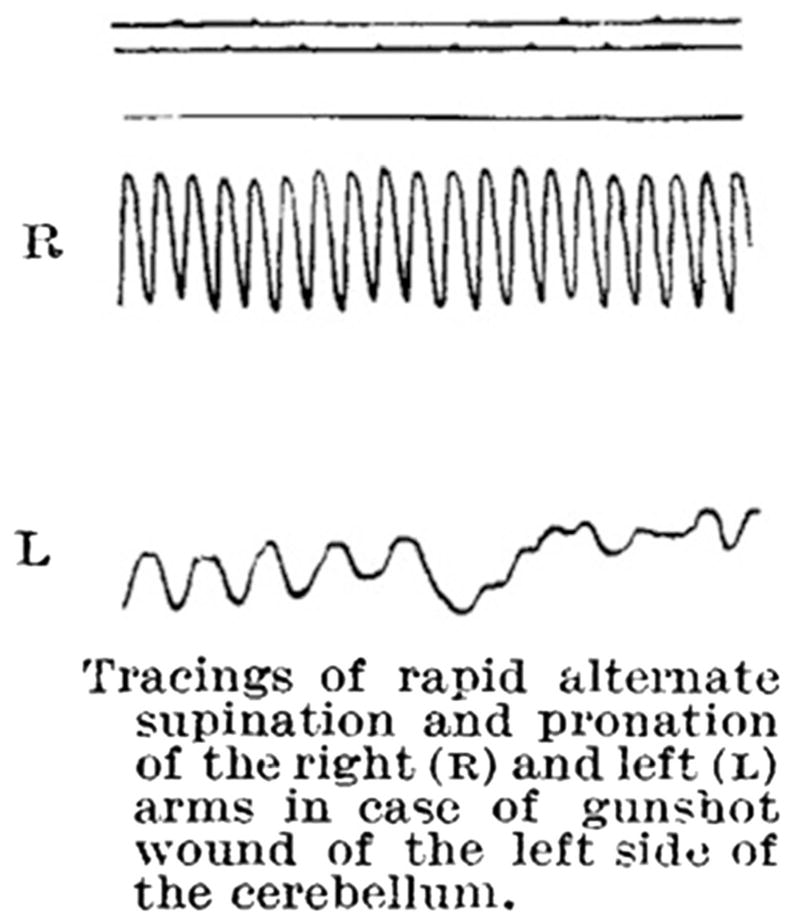
    1. Holmes G. The symptoms of acute cerebellar injuries due to gunshot injuries. Brain. 1917;40:461–535.
    1. Babinski J. Sur le role du cervelet dans les actes volitionnels nécessitant une succession rapide de mouvements (1) (diadococinésie) Rev Neurol. 1902;10:1013–5.

Publication types

MeSH terms