

Prenatal Nicotine Exposure Impairs the Proliferation of Neuronal Progenitors, Leading to Fewer Glutamatergic Neurons in the Medial Prefrontal Cortex
- PMID: 26105135
- PMCID: PMC5130133
- DOI: 10.1038/npp.2015.186
Prenatal Nicotine Exposure Impairs the Proliferation of Neuronal Progenitors, Leading to Fewer Glutamatergic Neurons in the Medial Prefrontal Cortex
Abstract
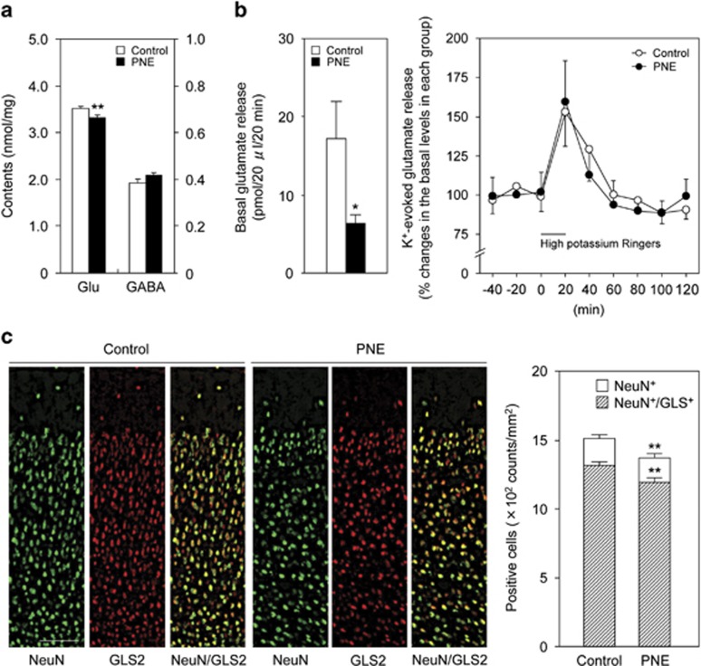
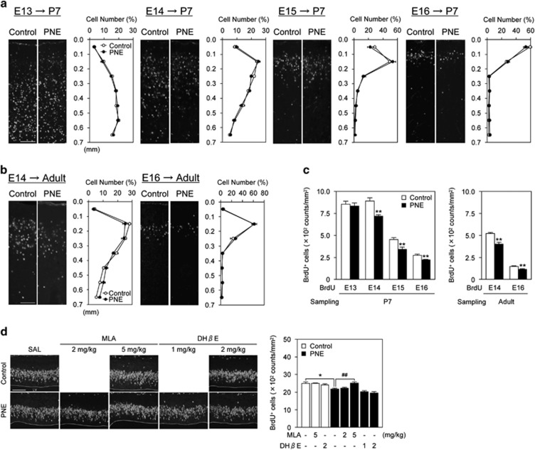
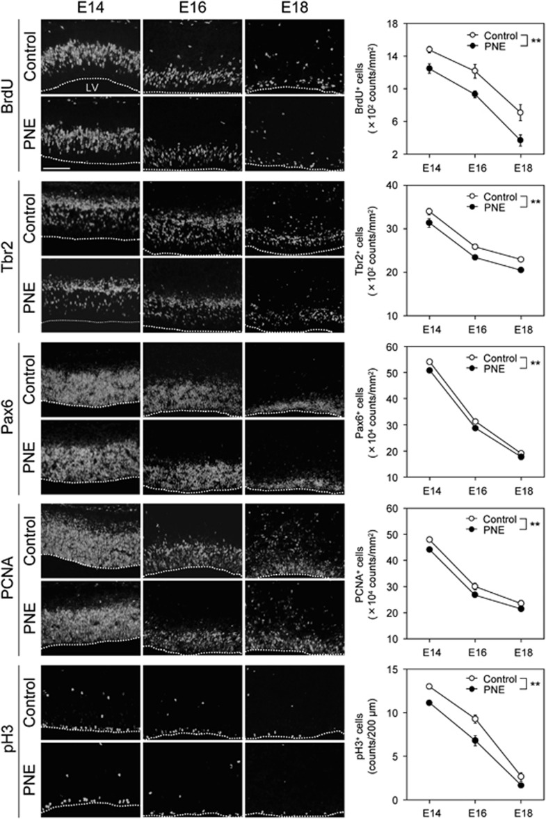
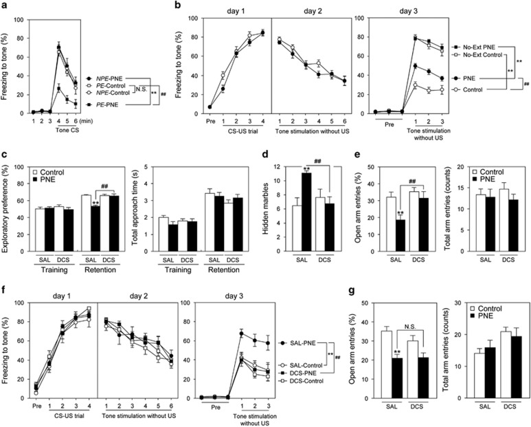
Cigarette smoking during pregnancy is associated with various disabilities in the offspring such as attention deficit/hyperactivity disorder, learning disabilities, and persistent anxiety. We have reported that nicotine exposure in female mice during pregnancy, in particular from embryonic day 14 (E14) to postnatal day 0 (P0), induces long-lasting behavioral deficits in offspring. However, the mechanism by which prenatal nicotine exposure (PNE) affects neurodevelopment, resulting in behavioral deficits, has remained unclear. Here, we report that PNE disrupted the proliferation of neuronal progenitors, leading to a decrease in the progenitor pool in the ventricular and subventricular zones. In addition, using a cumulative 5-bromo-2'-deoxyuridine labeling assay, we evaluated the rate of cell cycle progression causing the impairment of neuronal progenitor proliferation, and uncovered anomalous cell cycle kinetics in mice with PNE. Accordingly, the density of glutamatergic neurons in the medial prefrontal cortex (medial PFC) was reduced, implying glutamatergic dysregulation. Mice with PNE exhibited behavioral impairments in attentional function and behavioral flexibility in adulthood, and the deficits were ameliorated by microinjection of D-cycloserine into the PFC. Collectively, our findings suggest that PNE affects the proliferation and maturation of progenitor cells to glutamatergic neuron during neurodevelopment in the medial PFC, which may be associated with cognitive deficits in the offspring.
Figures
Similar articles
-
Prenatal Nicotine Exposure Impairs Executive Control Signals in Medial Prefrontal Cortex.Neuropsychopharmacology. 2016 Feb;41(3):716-25. doi: 10.1038/npp.2015.197. Epub 2015 Jul 20. Neuropsychopharmacology. 2016. PMID: 26189451 Free PMC article.
-
Prenatal NMDA receptor antagonism impaired proliferation of neuronal progenitor, leading to fewer glutamatergic neurons in the prefrontal cortex.Neuropsychopharmacology. 2012 May;37(6):1387-96. doi: 10.1038/npp.2011.324. Epub 2012 Jan 18. Neuropsychopharmacology. 2012. PMID: 22257896 Free PMC article.
-
Prenatal nicotine exposure decreases the release of dopamine in the medial frontal cortex and induces atomoxetine-responsive neurobehavioral deficits in mice.Psychopharmacology (Berl). 2017 Jun;234(12):1853-1869. doi: 10.1007/s00213-017-4591-z. Epub 2017 Mar 23. Psychopharmacology (Berl). 2017. PMID: 28332006
-
Behavioral and neural consequences of prenatal exposure to nicotine.J Am Acad Child Adolesc Psychiatry. 2001 Jun;40(6):630-41. doi: 10.1097/00004583-200106000-00007. J Am Acad Child Adolesc Psychiatry. 2001. PMID: 11392340 Review.
-
Prenatal Nicotine Exposure in Rodents: Why Are There So Many Variations in Behavioral Outcomes?Nicotine Tob Res. 2020 Oct 8;22(10):1694-1710. doi: 10.1093/ntr/ntz196. Nicotine Tob Res. 2020. PMID: 31595949 Review.
Cited by
-
Association between environmental tobacco smoke exposure across the first four years of life and manifestation of externalizing behavior problems in school-aged children.J Child Psychol Psychiatry. 2020 Nov;61(11):1243-1252. doi: 10.1111/jcpp.13157. Epub 2019 Dec 3. J Child Psychol Psychiatry. 2020. PMID: 31797389 Free PMC article.
-
Nicotine and the developing brain: Insights from preclinical models.Pharmacol Biochem Behav. 2022 Mar;214:173355. doi: 10.1016/j.pbb.2022.173355. Epub 2022 Feb 14. Pharmacol Biochem Behav. 2022. PMID: 35176350 Free PMC article.
-
Prenatal nicotine alters development of the laterodorsal tegmentum: Possible role for attention-deficit/hyperactivity disorder and drug dependence.World J Psychiatry. 2022 Feb 19;12(2):212-235. doi: 10.5498/wjp.v12.i2.212. eCollection 2022 Feb 19. World J Psychiatry. 2022. PMID: 35317337 Free PMC article. Review.
-
The cellular basis of fetal endoplasmic reticulum stress and oxidative stress in drug-induced neurodevelopmental deficits.Neurobiol Stress. 2018 Dec 27;10:100145. doi: 10.1016/j.ynstr.2018.100145. eCollection 2019 Feb. Neurobiol Stress. 2018. PMID: 30937351 Free PMC article. Review.
-
Transgenerational transmission of behavioral phenotypes produced by exposure of male mice to saccharin and nicotine.Sci Rep. 2020 Jul 20;10(1):11974. doi: 10.1038/s41598-020-68883-6. Sci Rep. 2020. PMID: 32686722 Free PMC article.
References
-
- Abreu-Villaca Y, Seidler FJ, Slotkin TA (2004). Does prenatal nicotine exposure sensitize the brain to nicotine-induced neurotoxicity in adolescence? Neuropsychopharmacology 29: 1440–1450. - PubMed
-
- Alkam T, Hiramatsu M, Mamiya T, Aoyama Y, Nitta A, Yamada K et al (2011). Evaluation of object-based attention in mice. Behav Brain Res 220: 185–193. - PubMed
-
- Alkam T, Kim HC, Hiramatsu M, Mamiya T, Aoyama Y, Nitta A et al (2013. a). Evaluation of emotional behaviors in young offspring of C57BL/6J mice after gestational and/or perinatal exposure to nicotine in six different time-windows. Behav Brain Res 239: 80–89. - PubMed
-
- Alkam T, Kim HC, Mamiya T, Yamada K, Hiramatsu M, Nabeshima T (2013. b). Evaluation of cognitive behaviors in young offspring of C57BL/6J mice after gestational nicotine exposure during different time-windows. Psychopharmacology (Berl) 230: 451–463. - PubMed
Publication types
MeSH terms
Substances
LinkOut - more resources
Full Text Sources
Other Literature Sources
Miscellaneous
