Review
. 2016 Jan;41(1):58-79.
doi: 10.1038/npp.2015.180. Epub 2015 Jun 24.

Stress and Fear Extinction

Affiliations
Review

Stress and Fear Extinction

Stephen Maren et al. Neuropsychopharmacology. 2016 Jan.

Abstract

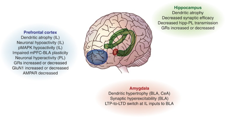
Stress has a critical role in the development and expression of many psychiatric disorders, and is a defining feature of posttraumatic stress disorder (PTSD). Stress also limits the efficacy of behavioral therapies aimed at limiting pathological fear, such as exposure therapy. Here we examine emerging evidence that stress impairs recovery from trauma by impairing fear extinction, a form of learning thought to underlie the suppression of trauma-related fear memories. We describe the major structural and functional abnormalities in brain regions that are particularly vulnerable to stress, including the amygdala, prefrontal cortex, and hippocampus, which may underlie stress-induced impairments in extinction. We also discuss some of the stress-induced neurochemical and molecular alterations in these brain regions that are associated with extinction deficits, and the potential for targeting these changes to prevent or reverse impaired extinction. A better understanding of the neurobiological basis of stress effects on extinction promises to yield novel approaches to improving therapeutic outcomes for PTSD and other anxiety and trauma-related disorders.


Figures

Figure 1
Figure 1
Relapse of fear after extinction. After the extinction of a conditioned fear response, extinction, the expression of conditioned fear is reduced. However, a number of phenomena are associated with the return or relapse of fear responses after extinction. These include (a) spontaneous recovery with the passage of time, (b) external disinhibition after presentation of a novel stimulus, (c) reinstatement after experiencing a noxious event, including the unconditioned stimulus, and (d) renewal after experiencing the conditioned stimulus outside the extinction context.
Figure 2
Figure 2
Stressors and stress models affecting fear extinction in rodents. A range of established stressors, applied singly or in combination, either acutely or chronically, has been shown to cause impairments in extinction acquisition or retrieval, sometimes in association with increased fear. Abnormalities in fear and extinction are also found after rodents are subjected to the stress-enhanced fear learning (SEFL) (Rau et al, 2005), single-prolonged stress (SPL) (Yamamoto et al, 2009), and immediate-extinction deficit (IED) (Maren, 2014b) models of stress.
Figure 3
Figure 3
Candidate gene variants moderating stress-related risk for posttraumatic stress disorder (PTSD). A number of human genetic variants have been found to interact with exposure to stress and trauma, often in childhood, to moderate risk for PTSD. To date, these have included the serotonin transporter (SL6A4) (Caspi et al, 2010; Xie et al, 2012), FK506-binding protein 51 (FKBP5) (Zannas and Binder, 2014), adenylate cyclase-activating polypeptide receptor (ADCYAP1) (Dias et al, 2013), and the β-adrenergic receptor (ADBR2) (Liberzon et al, 2014). Although they have not yet been studied for their potential interaction with stress, variants in the Tolloid-Like 1 Gene (TLL1) (Xie et al, 2013), retinoid-related orphan receptor alpha gene (RORA) (Logue et al, 2013) and fatty acid amide hydrolase (FAAH) (Dincheva, 2015; Gunduz-Cinar et al, 2013b) genes have been recently associated with PTSD or PTSD-related personality traits, including fear extinction and stress-reactivity.
Figure 4
Figure 4
Effects of stress on fear extinction in infant and adolescent rodents. (a) At preweaning ages, infant rodents exhibit an erasure-like form of extinction that is less prone to spontaneous recovery and context-renewal than extinction in post-weaning animals (Kim and Richardson, 2007). (b) Exposing infants to stressors leads to the emergence of the adult form of extinction at preweaning (Callaghan et al, 2013). (c) Adolescent rats exposed to chronic stress typically show impaired extinction when tested as adults (Ishikawa et al, 2012; Judo et al, 2010; Toledo-Rodriguez et al, 2012), although there is preliminary evidence that acute stress in adolescence can facilitate extinction (Schayek and Maroun, 2014).
Figure 5
Figure 5
Stress-induced changes in fear extinction-mediating brain regions. Stress produces a range of morphological, electrophysiological and molecular changes in the prefrontal cortex, hippocampus, and amygdala that parallel extinction abnormalities. BLA, basolateral nucleus of the amygdala; CeA, central nucleus of the amygdala; IL, infralimbic cortex; PL, prelimbic cortex.
Figure 6
Figure 6
Putative schematic flow of mechanism in the ventromedial prefrontal cortex (vmPFC) and basolateral nucleus of the amygdala (BLA) underlying stress-induced extinction deficits. In the vmPFC, stress has been shown to cause excess glutamate and associated dendritic hypotrophy and downregulation of glutamate receptors. These adaptations could lead to loss of vmPFC modulation of the amygdala and a resulting impairment in extinction that may be prevented by stimulating NMDA receptors using, eg, d-cycloserine. In the BLA, GR downregulation and anandamide depletion is seen after stress, which could in turn lead to reduced engagement of noradrenergic transmission and deficient extinction. There may be multiple points of intervention to reverse this cascade, including stimulating GRs or elevating anandamide with hydrocortisone and FAAH inhibitors, respectively, or blocking α2-adrenoceptors with yohimbine to increase BLA noradrenaline. BLA, basolateral amygdala; NE, norepinephrine; GR, glucocorticoid receptor; vmPFC, ventromedial prefrontal cortex.

Similar articles

Cited by

References

    1. Abrari K, Rashidy-Pour A, Semnanian S, Fathollahi Y (2008). Administration of corticosterone after memory reactivation disrupts subsequent retrieval of a contextual conditioned fear memory: dependence upon training intensity. Neurobiol Learn Mem 89: 178–184. - PubMed
    1. Andero R, Heldt SA, Ye K, Liu X, Armario A, Ressler KJ (2011). Effect of 7,8-dihydroxyflavone, a small-molecule TrkB agonist, on emotional learning. Am J Psychiatry 168: 163–172. - PMC - PubMed
    1. Atsak P, Hauer D, Campolongo P, Schelling G, Fornari RV, Roozendaal B (2014). Endocannabinoid signaling within the basolateral amygdala integrates multiple stress hormone effects on memory consolidation. Neuropsychopharmacology 40: 1485–1494. - PMC - PubMed
    1. Atsak P, Hauer D, Campolongo P, Schelling G, McGaugh JL, Roozendaal B (2012). Glucocorticoids interact with the hippocampal endocannabinoid system in impairing retrieval of contextual fear memory. Proc Natl Acad Sci USA 109: 3504–3509. - PMC - PubMed
    1. Auber A, Tedesco V, Jones CE, Monfils M-H, Chiamulera C (2013). Post-retrieval extinction as reconsolidation interference: methodological issues or boundary conditions? Psychopharmacology (Berl) 226: 631–647. - PMC - PubMed

Publication types

MeSH terms