

Long-term results of corneal collagen crosslinking for progressive keratoconus
- PMID: 26105541
- PMCID: PMC4502088
- DOI: 10.1016/j.optom.2014.05.006
Long-term results of corneal collagen crosslinking for progressive keratoconus
Abstract
Purpose: To evaluate long-term keratoconus stability after corneal crosslinking (CXL) with riboflavin.
Methods: In this prospective study, 57 eyes of 55 patients with progressive keratoconus, consecutively treated with ultraviolet A (UVA) - riboflavin CXL, were examined with the corneal topographer Pentacam, the biometer IOLMaster and the analyzer of corneal biomechanics Ocular Response Analyzer before and during a 24 months follow-up after CXL.
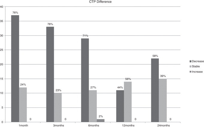
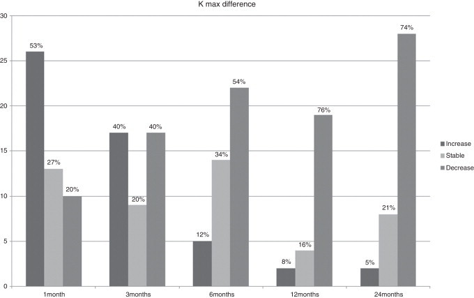
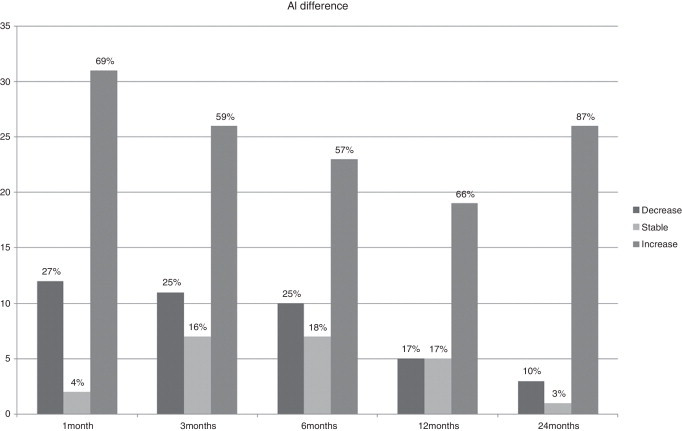
Results: Twenty-four months after CXL, there was a significant improvement in best corrected visual acuity (BCVA) (P<0.01), a significant decrease in corneal thinnest point (CTP), keratometry readings at the keratoconus apex (K max), and corneal volume (CV) (P<0.01), and a significant increase in axial eye length (AL) (P=0.01). No significant changes in anterior chamber volume (ACV) and depth (ACD), (P=0.8), corneal hysteresis (CH) (P=0.16) and corneal resistance factor (CRF) (P=0.06) were found. However, in the subgroup of patients with decreased K max readings 24 months after treatment, both CH and CRF showed a significant reduction (P<0.01).
Conclusion: In the first month after the procedure, CXL induces a reduction in corneal volume. During the 24 months follow-up the cornea tends to recover its original volume with a persistence of the CXL efficacy.
Objetivo: Evaluar la estabilidad del queratocono a largo plazo tras cross-linking corneal (CXL) con riboflavina.
Métodos: En este estudio prospectivo, se examinaron 57 ojos de 55 pacientes con queratocono progresivo, tratados consecutivamente con UVA-CXL con riboflavina utilizando el topógrafo corneal Pentacam, el biómetro IOLMaster y el analizar de la biomecánica corneal “Ocular Response Analyzer” preoperatoriamente y a los 24 meses de haberse realizado el CXL.
Resultados: A los veinticuatro meses del CXL, se produjo una mejora considerable de la agudeza visual mejor corregida (BCVA) (p < 0.01), un importante decremento del punto más fino de la córnea (CTP), de las medidas queratométricas en el vértice del queratocono (K max), y del volumen de la córnea (VC) (p < 0.01), y un incremento significativo de la longitud axial del ojo (LA) (p = 0.01). No se produjeron cambios significativos en el volumen de la cámara anterior (VCA) ni en la profundidad de la misma (PCA), (p = 0.8), histéresis de la córnea (HC) (p = 0.16) y factor de resistencia de la córnea (FRC) (p = 0.06). Sin embargo, en el subgrupo de pacientes con disminución de los valores de K max, a los 24 meses del tratamiento, tanto la HC como la FRC mostraron una reducción considerable (p < 0.01).
Conclusión: Durante el primer mes tras la intervención, el CXL induce una reducción del volumen de la córnea. Durante los 24 meses de seguimiento, la córnea tiende a recuperar su volumen original, persistiendo la eficacia del CXL.
Keywords: Axial eye length; Corneal crosslinking; Corneal thinnest point; Cross-linking corneal; Grosor corneal más delagado; Keratoconus; Keratometry readings; Longitud axial del ojo; Mediciones del queratómetro; Queratocono.
Copyright © 2013 Spanish General Council of Optometry. Published by Elsevier Espana. All rights reserved.
Figures
References
-
- Krachmer J.H., Feder R.S., Belin M.W. Keratoconus and related noninflammatory corneal thinning disorders. Surv Ophthalmol. 1984;28:293–322. - PubMed
-
- Tuft S.J., Moodaley L.C., Gregory W.M., Davison C.R., Buckley R.J. Prognostic factors for the progression of keratoconus. Ophthalmology. 1994;101:439–447. - PubMed
-
- Wollensak G., Spoerl E., Seiler T. Stress-strain measurements of human and porcine corneas after riboflavin-ultraviolet-A-induced cross-linking. J Cataract Refract Surg. 2003;29:1780–1785. - PubMed
-
- Kohlhaas M., Spoerl E., Schilde T., Unger G., Wittig C., Pillunat L.E. Biomechanical evidence of the distribution of cross-links in corneas treated with riboflavin and ultraviolet A light. J Cataract Refract Surg. 2006;32:279–283. - PubMed
MeSH terms
Substances
LinkOut - more resources
Full Text Sources
Other Literature Sources
Miscellaneous
