. 2015 May 22:9:22.
doi: 10.3389/fncir.2015.00022. eCollection 2015.

Functional response properties of VIP-expressing inhibitory neurons in mouse visual and auditory cortex

Affiliations

Functional response properties of VIP-expressing inhibitory neurons in mouse visual and auditory cortex

Lukas Mesik et al. Front Neural Circuits. .

Abstract

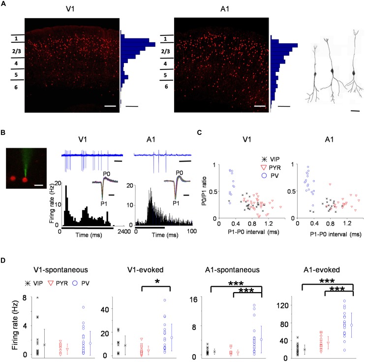
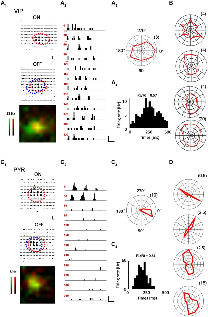
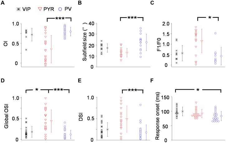
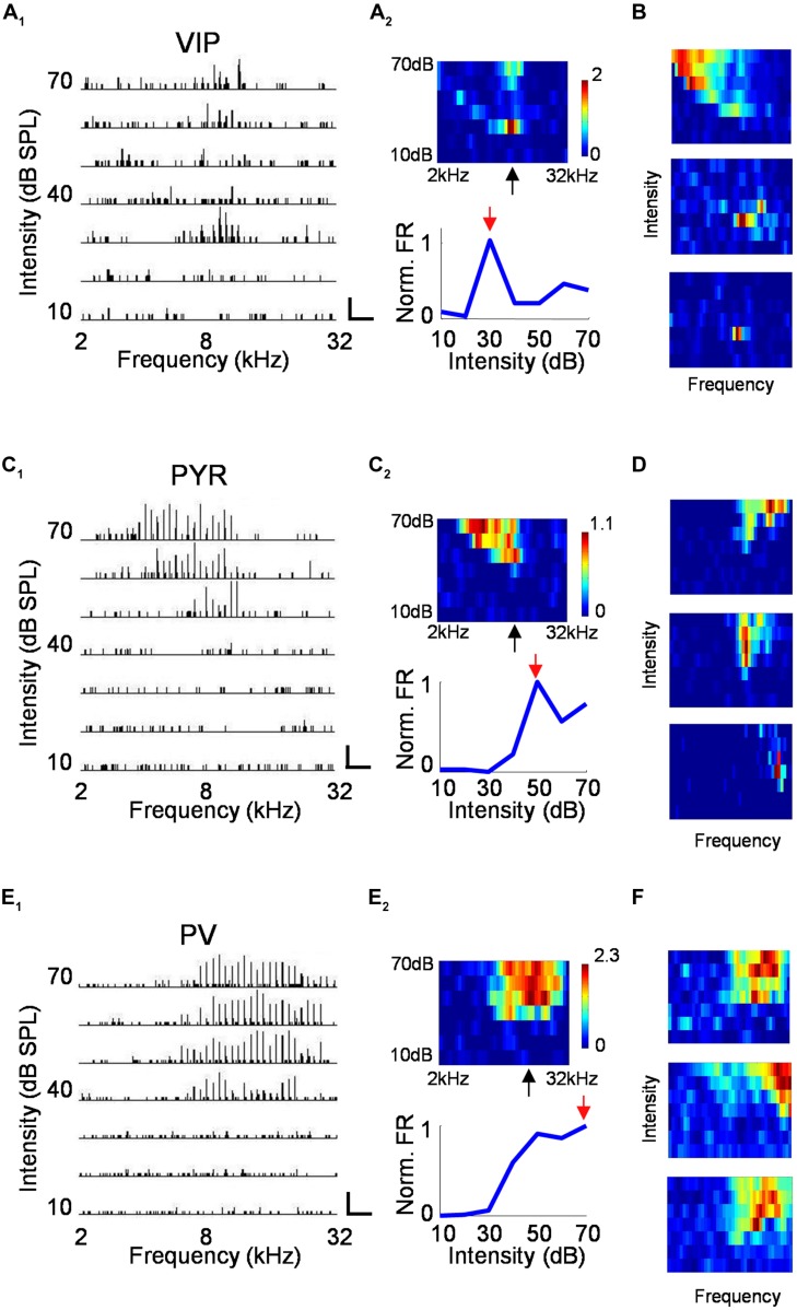
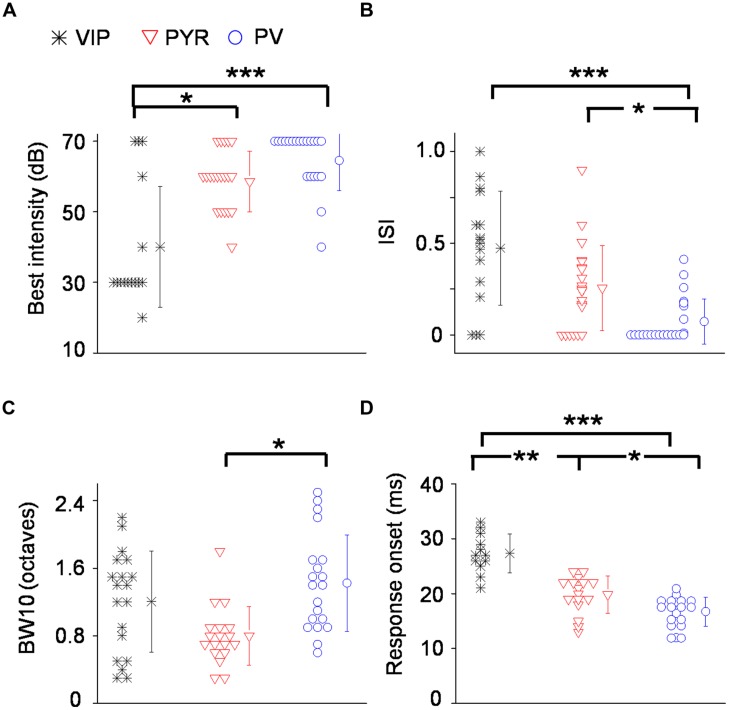
Despite accounting for about 20% of all the layer 2/3 inhibitory interneurons, the vasoactive intestinal polypeptide (VIP) expressing neurons remain the least thoroughly studied of the major inhibitory subtypes. In recent studies, VIP neurons have been shown to be activated by a variety of cortico-cortical and neuromodulatory inputs, but their basic sensory response properties remain poorly characterized. We set out to explore the functional properties of layer 2/3 VIP neurons in the primary visual (V1) and primary auditory cortex (A1), using two-photon imaging guided patch recordings. We found that in the V1, VIP neurons were generally broadly tuned, with their sensory response properties resembling those of parvalbumin (PV) expressing neurons. With the exception of response latency, they did not exhibit a significant difference from PV neurons across any of the properties tested, including overlap index, response modulation, orientation selectivity, and direction selectivity. In the A1, on the other hand, VIP neurons had a strong tendency to be intensity selective, which is a property associated with a subset of putative pyramidal cells and virtually absent in PV neurons. VIP neurons had a best intensity that was significantly lower than that of PV and putative pyramidal neurons. Finally, sensory evoked spike responses of VIP neurons were delayed relative to pyramidal and PV neurons in both the V1 and A1. Combined, these results demonstrate that the sensory response properties of VIP neurons do not fit a simple model of being either PV-like broadly tuned or pyramidal-like narrowly tuned. Instead, the selectivity pattern varies with sensory area and can even be, as in the case of low sound intensity responsiveness, distinct from both PV and pyramidal neurons.

Keywords: direction selectivity; frequency tuning; intensity selectivity; interneuron; orientation selectivity; receptive field property; tonal receptive field; visual receptive field.


Figures

FIGURE 1
FIGURE 1
Spiking properties of vasoactive intestinal polypeptide (VIP) neurons in the primary visual (V1) and primary auditory cortex (A1). (A) Coronal sections of a VIP-Cre::tdTomato brain showing red fluorescence labeled VIP neurons in the V1 (left) and A1 (right). Scale: 150 μm. Histograms adjacent to the images show the distribution of fluorescent cell bodies by depth within 50 μm bins. Scale: three cells per 50 μm × 00 μm × 100 μm. Far right, reconstructed morphologies of three representative VIP cells. Scale: 30 μm. (B) Left, a two-photon image showing a calcein-filled glass pipette forming a loose seal with a targeted VIP neuron. Scale: 20 μm. Middle, a sample recorded spike-response trace (2.4 s duration, scale: 300 ms; note that each vertical deflection reflects a spike current) of an example VIP neuron to a drifting grating (upper) and the post-stimulus spike time histogram (PSTH; lower) made for responses to all 12 orientations and of 5 repetitions. The black bar below indicates duration of the stimulus (onset at 0 s, 2 s long). Inset, superimposed individual spike waveforms during the recording for the displayed example cells. The trough and peak are labeled as P1 and P0, respectively. Scale: 1 ms. Right, tone-evoked responses of an example VIP neuron in the A1. The duration of the sample response trace is 100 ms (scale bar: 20 ms). The PSTH was made for responses to effective tones (38 tone–intensity combinations) and of nine repetitions. Black bar below indicates duration of the stimulus (onset at 0 s, 50 ms long). Scale for inset: 1 ms. (C) Scatterplots of P0/P1 amplitude ratio versus P1–P0 interval (a measure of spike width) for VIP (black), putative pyramidal (red) and PV (blue) cell populations. Each data point represents one cell. (D) Summaries of spontaneous and evoked firing rates of different cell populations. Mean values ± SD are shown for each cell group. Cell number: n = 20 in V1 and 17 in A1 for VIP; n = 22 in V1 and 22 in A1 for PV; 17 in V1 and 16 in A1 for Pyr. p < 0.05; ∗∗∗p < 0.001, Kruskal–Wallis (except for A1-evoked responses where ANOVA was used) and post hoc test.
FIGURE 2
FIGURE 2
Characterization of visual response properties. (A) An example VIP neuron. A1, array of PSTHs for the responses to 11 × 11 flash bright (On, upper) and dark (Off, lower) squares of 10 repetitions. The duration of flashing stimuli is indicated by a red bar. Scale: 12 Hz, 200 ms. Fitted On and Off response regions are marked by red and blue ovals, respectively. Bottom, superimposed color maps for On (red) and Off (green) responses. Color scale represents evoked firing rate. A2, PSTHs for responses to drifting sinusoidal gratings at 12 different directions. Scale: 10 Hz, 400 ms. A3, Polar graph plot of orientation-dependent response levels. The radius of the polar plot (firing rate in Hz) is indicated within parentheses. A4, Cycle averaged PSTH for spike responses evoked by drifting sinusoidal grating at optimal direction and spatial frequency. The modulation ratio F1/F0 is indicated. (B) Polar graph plots for four more example VIP neurons. (C) An example pyramidal neuron. Data are displayed in the same manner as in (A). Scale in C2: 33 Hz, 400 ms. (D) Polar graph plots for four more example pyramidal neurons.
FIGURE 3
FIGURE 3
Summary of visual response properties for VIP neurons as compared to PV and pyramidal neurons. (A) Distribution of overlap indices (OI). Mean values ± SD are shown for cell groups. N = 8 for VIP; 23 for Pyr; 25 for PV. (B) Distribution of subfield sizes. (C) Distribution of modulation (F1/F0) ratios. N = 10 for VIP; 18 for Pyr; 10 for PV. (D) Distribution of global orientation selectivity indices (gOSI). (E) Distribution of direction selectivity indices (DSI). (F) Distribution of spiking response onset latencies. N = 14 for VIP; 18 for Pyr; 19 for PV. p < 0.05; ∗∗∗p < 0.001, Kruskal–Wallis (except for B, where ANOVA was used) and post hoc test.
FIGURE 4
FIGURE 4
Characterization of auditory response properties. (A) An example VIP neuron. A1, tonal receptive field plotted as an array of PSTHs (five repetitions) to tones of varying intensity (10–70 dB SPL, 10 dB steps) and frequency (2–32 kHz, 0.1-octave steps). Scale: 29 Hz, 500 ms. Tone duration was 100 ms. A2, top, color map representation of the tonal receptive field (TRF). Pixel color represents the average number of spikes evoked per stimulus repetition within a 100-ms analysis window. Black arrow points to the characteristic frequency (CF). Bottom, response levels along the intensity, measured at and close to the CF and normalized by the maximum response level. Red arrow points to the best intensity. (B) Color maps for TRFs of another three example VIP neurons. Color scale (from top to bottom): 3.4; 1.3; 0.9 for maximum. (C,D) Example pyramidal neurons. Data are presented in the same manner as in (A,B). Scale in C1: 50 Hz, 250 ms. Color scale for (D; from top to bottom): 2.8; 2.2; 1.8 for maximum. (E,F) Example PV neurons. Scale in E1: 75 Hz, 250 ms. Color scale for (D; from top to bottom): 4.2; 3.5; 2.8 for maximum.
FIGURE 5
FIGURE 5
Summary of auditory response properties of VIP neurons as compared to PV and pyramidal neurons. (A) Distribution of best intensities. N = 16 for VIP; 21 for Pyr; 22 for PV. (B) Distribution of intensity selectivity indices (ISI). (C) Distribution of TRF bandwidths (BWs) measured at 10 dB above intensity threshold (or at the threshold). (D) Distribution of spiking response onset latencies. p < 0.05; ∗∗p < 0.01; ∗∗∗p < 0.001, Kruskal–Wallis and post hoc test.
FIGURE 6
FIGURE 6
Spontaneous firing rates during recording sessions. (A) Time course of average spontaneous firing rates for a VIP neuron in the A1 (left) and a VIP neuron in the V1 (right). Each data point is the measure of average spontaneous firing rate during the receptive field mapping of one repetition. (B) Average firing rates for the first and second half duration of the recording session for all the VIP cells recorded in the A1. (C) Average firing rates for the first and second half duration of the recording session for all the VIP cells recorded in the V1. (D) Average firing rates for the first and second half duration of the recording session for all the PV cells recorded in the A1. (D) Average firing rates for the first and second half duration of the recording session for all the putative pyramidal cells recorded in the A1. n.s., non-significant, paired t-test.

Similar articles

Cited by

References

    1. Adesnik H., Bruns W., Taniguchi H., Huang Z. J., Scanziani M. (2012). A neural circuit for spatial summation in visual cortex. Nature 490 226–231. 10.1038/nature11526 - DOI - PMC - PubMed
    1. Alitto H. J., Dan Y. (2012). Cell-type-specific modulation of neocortical activity by basal forebrain input. Front. Sys. Neurosci. 6:79 10.3389/fnsys.2012.00079 - DOI - PMC - PubMed
    1. Arroyo S., Bennett C., Aziz D., Brown S. P., Hestrin S. (2012). Prolonged disynaptic inhibition in the cortex mediated by slow, non-α7 nicotinic excitation of a specific subset of cortical interneurons. J. Neurosci. 32 3859–3864. 10.1523/JNEUROSCI.0115-12.2012 - DOI - PMC - PubMed
    1. Ascoli G. A., Alonso-Nanclares L., Anderson S. A., Barrionuevo G., Benavides-Piccione R., Burkhalter A., et al. (2008). Petilla terminology: nomenclature of features of GABAergic interneurons of the cerebral cortex. Nat. Rev. Neurosci. 9 557–568. 10.1038/nrn2402 - DOI - PMC - PubMed
    1. Atallah B. V., Bruns W., Carandini M., Scanziani M. (2012). Parvalbumin-expressing interneurons linearly transform cortical responses to visual stimuli. Neuron 73 159–170. 10.1016/j.neuron.2011.12.013 - DOI - PMC - PubMed

Publication types

MeSH terms

Substances

LinkOut - more resources