

Changes in task-based effective connectivity in language networks following rehabilitation in post-stroke patients with aphasia
- PMID: 26106314
- PMCID: PMC4460429
- DOI: 10.3389/fnhum.2015.00316
Changes in task-based effective connectivity in language networks following rehabilitation in post-stroke patients with aphasia
Abstract
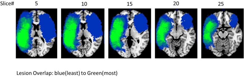
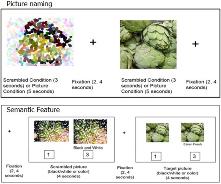
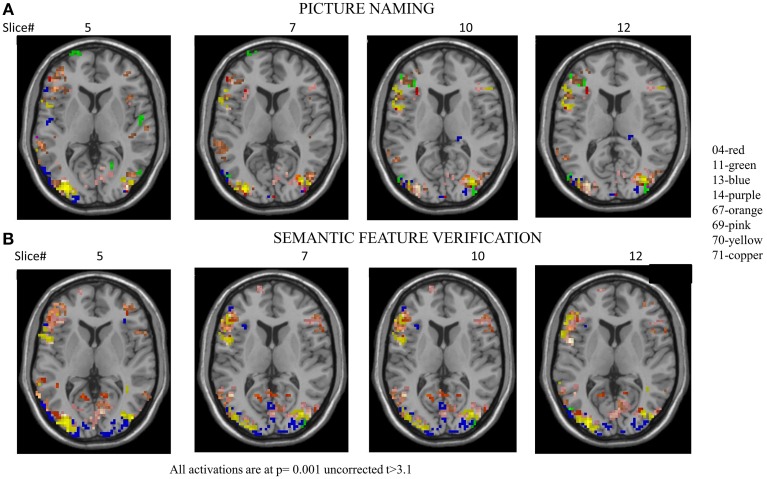
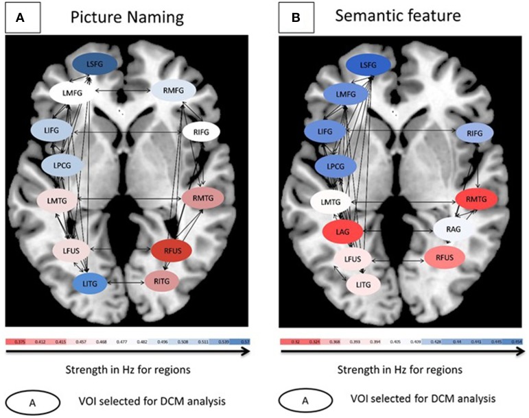
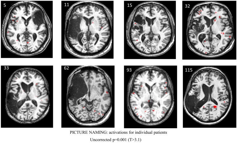
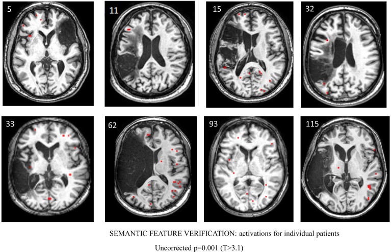
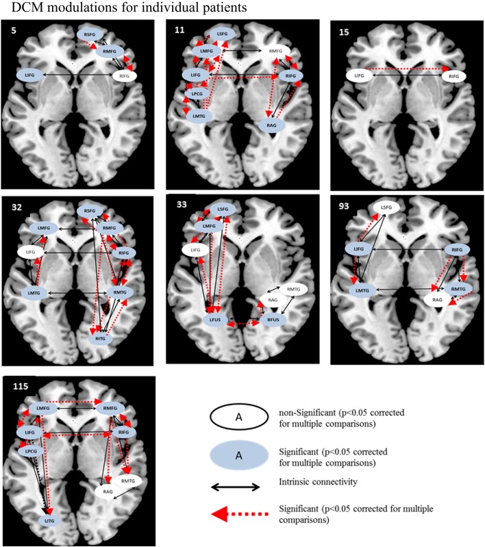
In this study, we examined regions in the left and right hemisphere language network that were altered in terms of the underlying neural activation and effective connectivity subsequent to language rehabilitation. Eight persons with chronic post-stroke aphasia and eight normal controls participated in the current study. Patients received a 10 week semantic feature-based rehabilitation program to improve their skills. Therapy was provided on atypical examples of one trained category while two control categories were monitored; the categories were counterbalanced across patients. In each fMRI session, two experimental tasks were conducted: (a) picture naming and (b) semantic feature verification of trained and untrained categories. Analysis of treatment effect sizes revealed that all patients showed greater improvements on the trained category relative to untrained categories. Results from this study show remarkable patterns of consistency despite the inherent variability in lesion size and activation patterns across patients. Across patients, activation that emerged as a function of rehabilitation on the trained category included bilateral IFG, bilateral SFG, LMFG, and LPCG for picture naming; and bilateral IFG, bilateral MFG, LSFG, and bilateral MTG for semantic feature verification. Analysis of effective connectivity using Dynamic Causal Modeling (DCM) indicated that LIFG was the consistently significantly modulated region after rehabilitation across participants. These results indicate that language networks in patients with aphasia resemble normal language control networks and that this similarity is accentuated by rehabilitation.
Keywords: aphasia; dynamic causal modeling; effective connectivity; fMRI activations; language recovery; naming; rehabilitation; stroke.
Figures

Similar articles
-
The Relationship between Frontotemporal Effective Connectivity during Picture Naming, Behavior, and Preserved Cortical Tissue in Chronic Aphasia.Front Hum Neurosci. 2016 Mar 16;10:109. doi: 10.3389/fnhum.2016.00109. eCollection 2016. Front Hum Neurosci. 2016. PMID: 27014039 Free PMC article.
-
Treatment-related changes in neural activation vary according to treatment response and extent of spared tissue in patients with chronic aphasia.Cortex. 2019 Dec;121:147-168. doi: 10.1016/j.cortex.2019.08.016. Epub 2019 Sep 19. Cortex. 2019. PMID: 31627014 Free PMC article.
-
Dynamic reorganization of task-related network interactions in post-stroke aphasia recovery.Brain. 2025 Jan 30:awaf036. doi: 10.1093/brain/awaf036. Online ahead of print. Brain. 2025. PMID: 39883566
-
Resting-state functional connectivity: An emerging method for the study of language networks in post-stroke aphasia.Brain Cogn. 2019 Apr;131:22-33. doi: 10.1016/j.bandc.2017.08.005. Epub 2017 Aug 31. Brain Cogn. 2019. PMID: 28865994 Review.
-
Remapping and Reconnecting the Language Network after Stroke.Brain Sci. 2024 Apr 24;14(5):419. doi: 10.3390/brainsci14050419. Brain Sci. 2024. PMID: 38790398 Free PMC article. Review.
Cited by
-
Altered brain language network in idiopathic peripheral facial paralysis patients with dysarthria.Ann Transl Med. 2020 Jun;8(11):699. doi: 10.21037/atm.2020.03.133. Ann Transl Med. 2020. PMID: 32617319 Free PMC article.
-
Pre-treatment graph measures of a functional semantic network are associated with naming therapy outcomes in chronic aphasia.Brain Lang. 2020 Aug;207:104809. doi: 10.1016/j.bandl.2020.104809. Epub 2020 Jun 5. Brain Lang. 2020. PMID: 32505940 Free PMC article.
-
Right Hemisphere Grey Matter Volume and Language Functions in Stroke Aphasia.Neural Plast. 2017;2017:5601509. doi: 10.1155/2017/5601509. Epub 2017 May 9. Neural Plast. 2017. PMID: 28573050 Free PMC article.
-
Disentangling neuroplasticity mechanisms in post-stroke language recovery.Brain Lang. 2024 Apr;251:105381. doi: 10.1016/j.bandl.2024.105381. Epub 2024 Feb 23. Brain Lang. 2024. PMID: 38401381 Free PMC article. Review.
-
Neural mechanisms of sentence production: a volumetric study of primary progressive aphasia.Cereb Cortex. 2024 Jan 14;34(1):bhad470. doi: 10.1093/cercor/bhad470. Cereb Cortex. 2024. PMID: 38100360 Free PMC article.
References
Grants and funding
LinkOut - more resources
Full Text Sources
Other Literature Sources
Medical