. 2015 Jun 9:6:543.
doi: 10.3389/fmicb.2015.00543. eCollection 2015.

S-layers at second glance? Altiarchaeal grappling hooks (hami) resemble archaeal S-layer proteins in structure and sequence

Affiliations

S-layers at second glance? Altiarchaeal grappling hooks (hami) resemble archaeal S-layer proteins in structure and sequence

Alexandra K Perras et al. Front Microbiol. .

Abstract

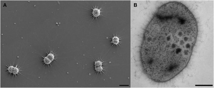
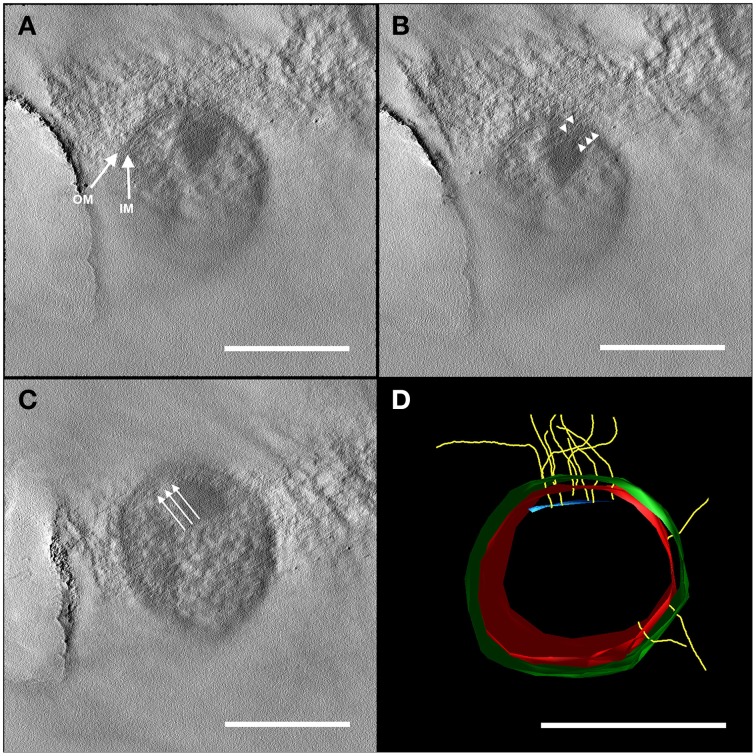
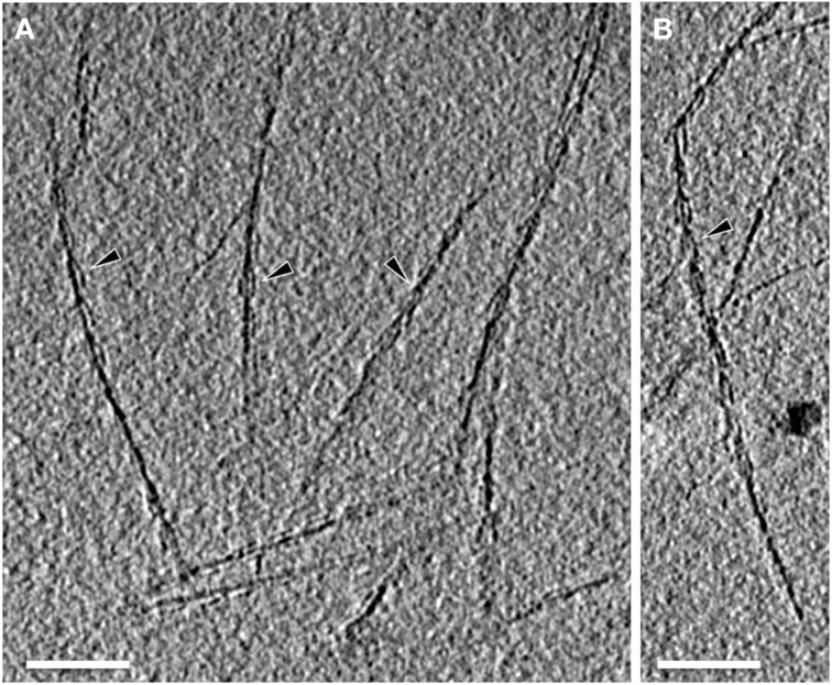
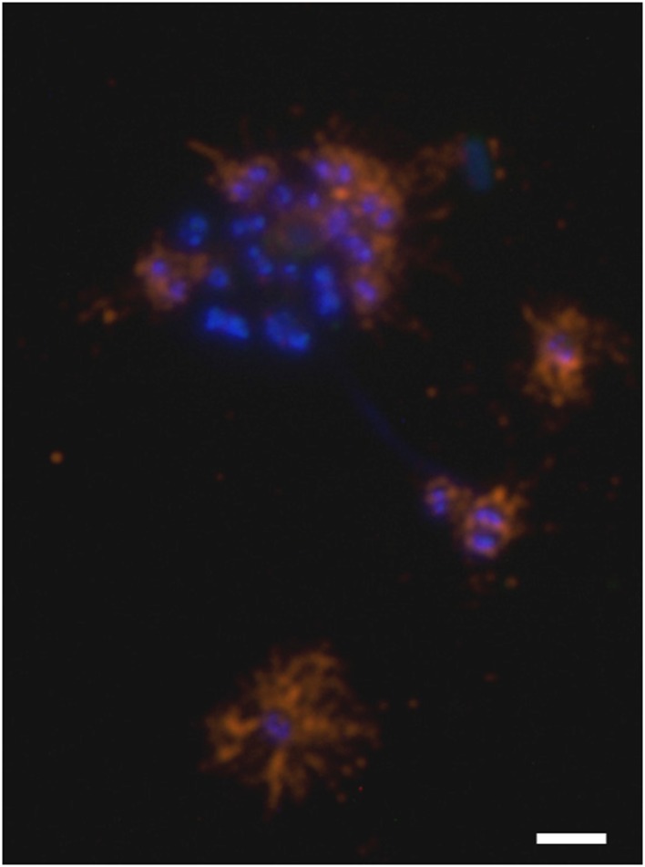
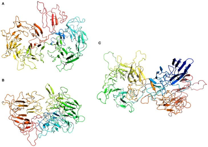
The uncultivated "Candidatus Altiarchaeum hamiconexum" (formerly known as SM1 Euryarchaeon) carries highly specialized nano-grappling hooks ("hami") on its cell surface. Until now little is known about the major protein forming these structured fibrous cell surface appendages, the genes involved or membrane anchoring of these filaments. These aspects were analyzed in depth in this study using environmental transcriptomics combined with imaging methods. Since a laboratory culture of this archaeon is not yet available, natural biofilm samples with high Ca. A. hamiconexum abundance were used for the entire analyses. The filamentous surface appendages spanned both membranes of the cell, which are composed of glycosyl-archaeol. The hami consisted of multiple copies of the same protein, the corresponding gene of which was identified via metagenome-mapped transcriptome analysis. The hamus subunit proteins, which are likely to self-assemble due to their predicted beta sheet topology, revealed no similiarity to known microbial flagella-, archaella-, fimbriae- or pili-proteins, but a high similarity to known S-layer proteins of the archaeal domain at their N-terminal region (44-47% identity). Our results provide new insights into the structure of the unique hami and their major protein and indicate their divergent evolution with S-layer proteins.

Keywords: S-layers; archaea; archaeal cell surface appendages; double-membrane; electron cryo-tomography; environmental transcriptomics; hami; nano-grappling hooks.


Figures

Figure 1
Figure 1
Electron micrographs of Ca. A. hamiconexum cells within the biofilm. (A) Scanning electron micrograph of dividing cells in the biofilm, showing particles close to the hami and the cells. Bar: 1 μm. (B) Thin-section of one cell, showing dark-stained areas. Bar. 200 nm.
Figure 2
Figure 2
Electron tomography of a 50 nm section of high-pressure frozen and freeze substituted cells. The tilt series was carried out at 120 kV from −60 to +60° with an increment of 2° and a final magnification of 14,000 x. The reconstruction of the tomogram was performed with IMOD. In selected virtual slices, several structures were indicated. (A) The final tomogram revealed the inner (IM) and outer membrane (OM) with a thickness of about 6 nm in each case. The overall thickness of inner and OM together with the periplasm was 44 nm. Therefore, the periplasm has a width of about 32 nm. (B) Further, two hami filaments with a diameter of approximately 8 nm were indicated spanning both the IM and OM (arrow heads). (C) Underneath the IM, another layer could be recognized (arrows). As the resolution of the tomogram is quite low, this could either be kind of an anchoring structure/mechano sensor or a preparation/reconstruction artifact as it was just visible in a very low number of the virtual slices of the tomogram. With this tomogram, a model (segmentation) was constructed (D) illustrating the IM (red) and OM (green), the hami filaments (yellow) and the supposed anchoring-associated structure (blue). For simplicity, the membranes were illustrated as a monolayer. Scale bars: 500 nm.
Figure 3
Figure 3
Electron cryo-tomography of the hami. Tomographic slices through hami emanating from the cell surface. Hami occur as single barbed wire-like filaments (A,B; black arrowheads). Scale bar: 100 nm.
Figure 4
Figure 4
Electron micrographs of isolated hami. (A,B) Overview of negatively stained hami with visible triple grappling hooks. The typical barbed-wire structure of the filament is shown, next to plain (probably) stretched filaments. Electron cryo-tomography shows the bundling of the hami (C, white arrow). Scale bar: 500 nm.
Figure 5
Figure 5
Immuno-staining of cells embedded in the archaeal biofilm. DNA is stained blue (DAPI) and hami were visualized using a CY3-labeled anti-hamus antibody (orange). The hami formed a halo around the cell. Scale bar: 2 μm.
Figure 6
Figure 6
Homology modeling of the Hami sequence on (A) a cytoplasmic response regulator of two-component system (pdb entry 4a2l), (B) a xyloglucanase from Clostridium thermocellum (pdb entry 2cn3), and (C) a human DNA-damage binding protein (pdb entry 3ei3). The models are displayed as cartoons colored in spectrum mode with the N-terminal region in blue and the C-terminal region in red.

References

    1. Albers S.-V., Pohlschröder M. (2009). Diversity of archaeal type IV pilin-like structures. Extremophiles 13, 403–410. 10.1007/s00792-009-0241-7 - DOI - PubMed
    1. Altschul S. F., Gish W., Miller W., Myers E. W., Lipman D. J. (1990). Basic local alignment search tool. J. Mol. Biol. 215, 403–410. 10.1016/S0022-2836(05)80360-2 - DOI - PubMed
    1. Arbing M. A., Chan S., Shin A., Phan T., Ahn C. J., Rohlin L., et al. . (2012). Structure of the surface layer of the methanogenic archaean Methanosarcina acetivorans. Proc. Natl. Acad. Sci. U.S.A. 109, 11812–11817. 10.1073/pnas.1120595109 - DOI - PMC - PubMed
    1. Arts J., van Boxtel R., Filloux A., Tommassen J., Koster M. (2007). Export of the pseudopilin XcpT of the Pseudomonas aeruginosa type II secretion system via the signal recognition particle-Sec pathway. J. Bacteriol. 189, 2069–2076. 10.1128/JB.01236-06 - DOI - PMC - PubMed
    1. Ayoub N. A., Garb J. E., Tinghitella R. M., Collin M. A., Hayashi C. Y. (2007). Blueprint for a high-performance biomaterial: full-length spider dragline silk genes. PLoS ONE 2:e514. 10.1371/journal.pone.0000514 - DOI - PMC - PubMed