

Automated quantification of the phagocytosis of Aspergillus fumigatus conidia by a novel image analysis algorithm
- PMID: 26106370
- PMCID: PMC4460560
- DOI: 10.3389/fmicb.2015.00549
Automated quantification of the phagocytosis of Aspergillus fumigatus conidia by a novel image analysis algorithm
Abstract
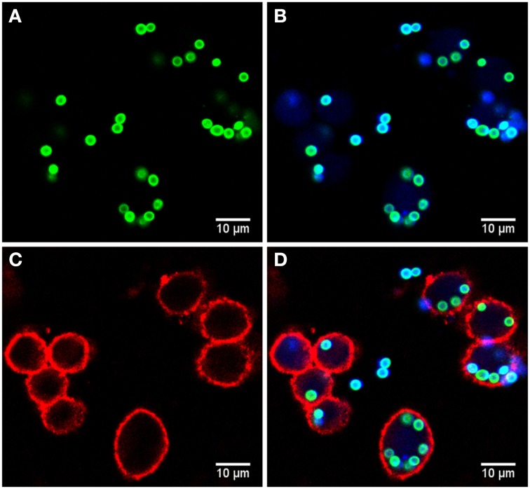
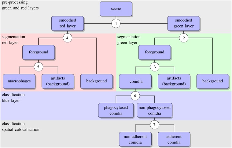
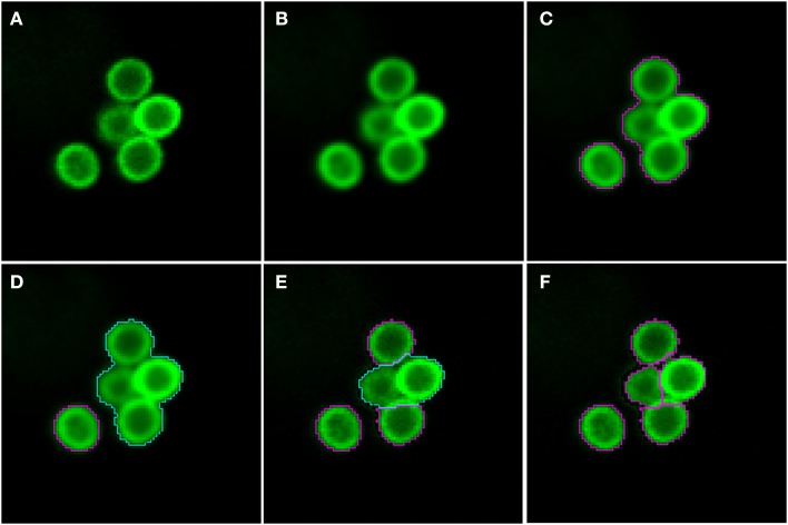
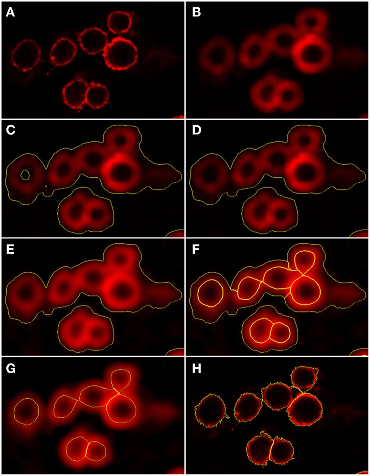
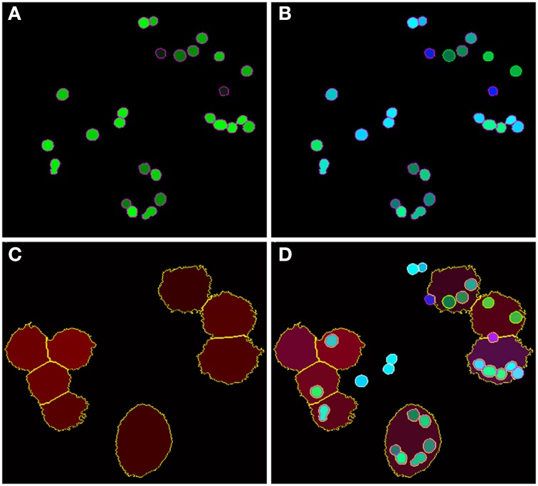
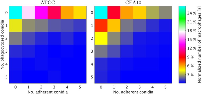
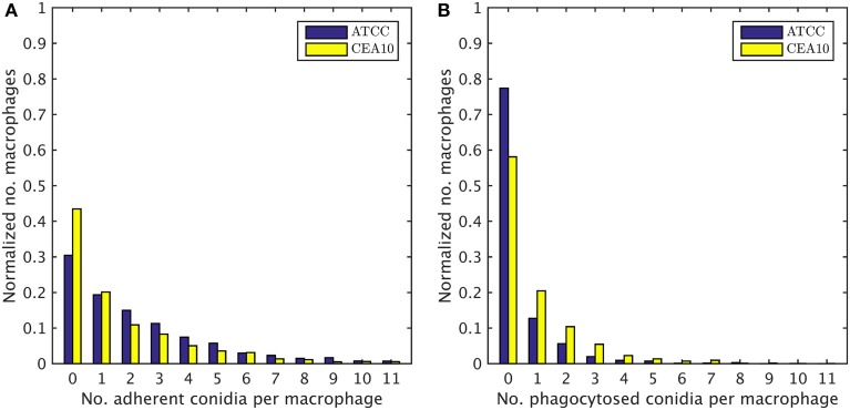
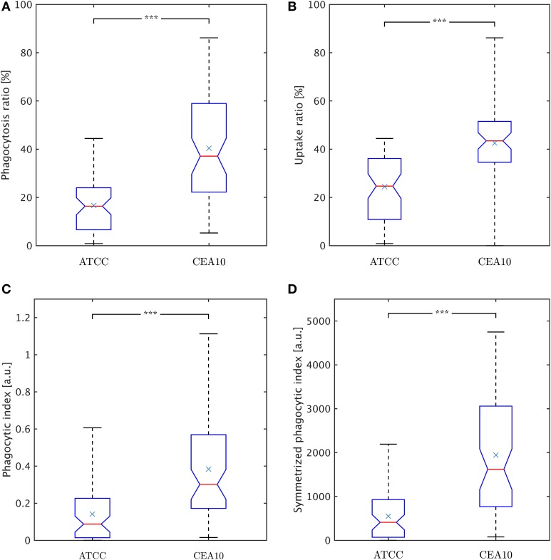
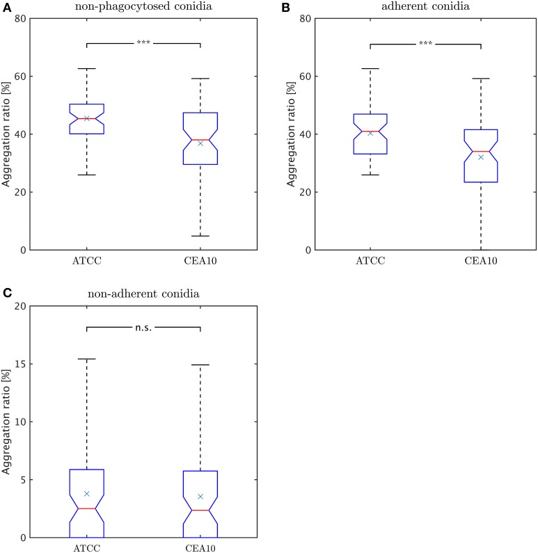
Studying the pathobiology of the fungus Aspergillus fumigatus has gained a lot of attention in recent years. This is due to the fact that this fungus is a human pathogen that can cause severe diseases, like invasive pulmonary aspergillosis in immunocompromised patients. Because alveolar macrophages belong to the first line of defense against the fungus, here, we conduct an image-based study on the host-pathogen interaction between murine alveolar macrophages and A. fumigatus. This is achieved by an automated image analysis approach that uses a combination of thresholding, watershed segmentation and feature-based object classification. In contrast to previous approaches, our algorithm allows for the segmentation of individual macrophages in the images and this enables us to compute the distribution of phagocytosed and macrophage-adherent conidia over all macrophages. The novel automated image-based analysis provides access to all cell-cell interactions in the assay and thereby represents a framework that enables comprehensive computation of diverse characteristic parameters and comparative investigation for different strains. We here apply automated image analysis to confocal laser scanning microscopy images of the two wild-type strains ATCC 46645 and CEA10 of A. fumigatus and investigate the ability of macrophages to phagocytose the respective conidia. It is found that the CEA10 strain triggers a stronger response of the macrophages as revealed by a higher phagocytosis ratio and a larger portion of the macrophages being active in the phagocytosis process.
Keywords: Aspergillus fumigatus; alveolar macrophages; automated image analysis; host-pathogen interaction; phagocytosis assay.
Figures
Similar articles
-
Automated image analysis of the host-pathogen interaction between phagocytes and Aspergillus fumigatus.PLoS One. 2011 May 5;6(5):e19591. doi: 10.1371/journal.pone.0019591. PLoS One. 2011. PMID: 21573171 Free PMC article.
-
Endocytic Markers Associated with the Internalization and Processing of Aspergillus fumigatus Conidia by BEAS-2B Cells.mSphere. 2019 Feb 6;4(1):e00663-18. doi: 10.1128/mSphere.00663-18. mSphere. 2019. PMID: 30728282 Free PMC article.
-
Phagocytosis and intracellular fate of Aspergillus fumigatus conidia in alveolar macrophages.Infect Immun. 2003 Feb;71(2):891-903. doi: 10.1128/IAI.71.2.891-903.2003. Infect Immun. 2003. PMID: 12540571 Free PMC article.
-
Pulmonary defense mechanisms against opportunistic fungal pathogens.Immunol Ser. 1989;47:243-71. Immunol Ser. 1989. PMID: 2490078 Review.
-
Intracellular and extracellular growth of Aspergillus fumigatus.Med Mycol. 2005 May;43 Suppl 1:S27-30. doi: 10.1080/13693780400029247. Med Mycol. 2005. PMID: 16110789 Review.
Cited by
-
Targeting of phagolysosomes containing conidia of the fungus Aspergillus fumigatus with polymeric particles.Appl Microbiol Biotechnol. 2023 Feb;107(2-3):819-834. doi: 10.1007/s00253-022-12287-1. Epub 2022 Dec 8. Appl Microbiol Biotechnol. 2023. PMID: 36480041 Free PMC article.
-
On the lineage of Aspergillus fumigatus isolates in common laboratory use.Med Mycol. 2021 Jan 4;59(1):7-13. doi: 10.1093/mmy/myaa075. Med Mycol. 2021. PMID: 32944768 Free PMC article.
-
A Multiparametric and High-Throughput Assay to Quantify the Influence of Target Size on Phagocytosis.Biophys J. 2019 Aug 6;117(3):408-419. doi: 10.1016/j.bpj.2019.06.021. Epub 2019 Jun 26. Biophys J. 2019. PMID: 31301802 Free PMC article.
-
Characterisation of Aspergillus fumigatus Endocytic Trafficking within Airway Epithelial Cells Using High-Resolution Automated Quantitative Confocal Microscopy.J Fungi (Basel). 2021 Jun 7;7(6):454. doi: 10.3390/jof7060454. J Fungi (Basel). 2021. PMID: 34200399 Free PMC article.
-
Quantitative Impact of Cell Membrane Fluorescence Labeling on Phagocytosis Measurements in Confrontation Assays.Front Microbiol. 2020 Jun 5;11:1193. doi: 10.3389/fmicb.2020.01193. eCollection 2020. Front Microbiol. 2020. PMID: 32582113 Free PMC article.
References
-
- Blinchikoff H., Krause H. (2001). Filtering in the Time and Frequency Domains. London: The Institution of Engineering and Technology.
-
- Brakhage A. A., Jahn B., Schmidt A. (eds.). (1999). Aspergillus Fumigatus: Biology, Clinical Aspects, and Molecular Approaches to Pathogenicity, Vol. 2. Basel: Karger Medical and Scientific Publishers.
LinkOut - more resources
Full Text Sources
Other Literature Sources