. 2015 Feb 9:8:314-21.
doi: 10.1016/j.nicl.2015.02.001. eCollection 2015.

Cerebellar infarct patterns: The SMART-Medea study

Collaborators, Affiliations

Cerebellar infarct patterns: The SMART-Medea study

Laurens J L De Cocker et al. Neuroimage Clin. .

Abstract

Objective: Previous studies on cerebellar infarcts have been largely restricted to acute infarcts in patients with clinical symptoms, and cerebellar infarcts have been evaluated with the almost exclusive use of transversal MR images. We aimed to document the occurrence and 3D-imaging patterns of cerebellar infarcts presenting as an incidental finding on MRI.

Methods: We analysed the 1.5 Tesla MRI, including 3D T1-weighted datasets, of 636 patients (mean age 62 ± 9 years, 81% male) from the SMART-Medea study. Cerebellar infarct analyses included an assessment of size, cavitation and gliosis, of grey and white matter involvement, and of infarct topography.

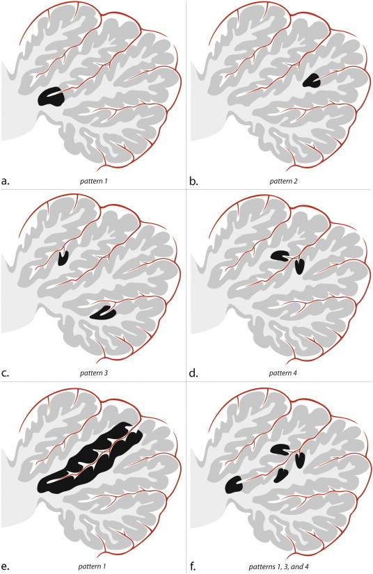
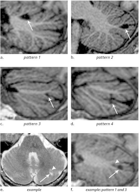
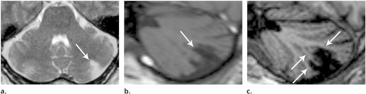
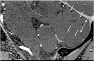
Results: One or more cerebellar infarcts (mean 1.97; range 1-11) were detected in 70 out of 636 patients (11%), with a total amount of 138 infarcts identified, 135 of which showed evidence of cavitation. The average mean axial diameter was 7 mm (range 2-54 mm), and 131 infarcts (95%) were smaller than 20 mm. Hundred-thirty-four infarcts (97%) involved the cortex, of which 12 in combination with subcortical white matter. No infarcts were restricted to subcortical branches of white matter. Small cortical infarcts involved the apex of a deep (pattern 1) or shallow fissure (pattern 2), or occurred alongside one (pattern 3) or opposite sides (pattern 4) of a fissure. Most (87%) cerebellar infarcts were situated in the posterior lobe.

Conclusions: Small cerebellar infarcts proved to be much more common than larger infarcts, and preferentially involved the cortex. Small cortical infarcts predominantly involved the posterior lobes, showed sparing of subcortical white matter and occurred in characteristic topographic patterns.

Keywords: Cerebellum; Cerebrovascular disease; MRI.


Figures

Fig. 1
Fig. 1
Schematic sagittal drawings illustrate a single cerebellar lobe, its arterial supply, and the patterns of small infarcts. Within the depicted cerebellar lobe, multiple folia are separated by fissures. The folia, which consist of the cortex and subcortical white matter, converge towards the deep white matter of the cerebellum. In the fissures, an arterial branch is present which gives rise to cortical arteries. (a) Pattern 1 corresponds to infarcts involving the apex of a large fissure, (b) pattern 2 corresponds to infarcts involving the apex of a shallow fissure, (c) two infarcts corresponding to pattern 3, and (d) one infarct involving opposite sides of a fissure, indicative of pattern 4. (e) Infarct involving the entire cortical coating of a deep cerebellar fissure; notice this also is a pattern 1 infarct since it involves the apex of a deep fissure. This infarct likely resulted from the occlusion of the arterial branch in the cerebellar fissure. (f) Combinations of small infarcts commonly occur alongside the same fissure. (a–f) Notice the sparing of both subcortical and deep white matter in each cortical infarct.
Fig. 2
Fig. 2
(a–d) 3D T1-weighted images (sagittal reconstructions) of the brain, providing excellent contrast between grey matter and white matter, show the four patterns by which cerebellar infarcts (arrows) typically affect the cerebellar cortex. (e) Example of two cerebellar infarcts adjacent to each other (arrow and arrowhead), detected on transverse T2WI. (f) Sagittal T1-weighted image of the same two infarcts shows how the topography of one infarct corresponds to pattern 3 (arrow), while the other infarct (arrowhead) corresponds to a small pattern 1 infarct. Notice the sparing of the subcortical and deep white matter in each infarct. Images are cropped to display the cerebellum only.
Fig. 3
Fig. 3
Although this larger infarct spans multiple fissures, it is seen to spare the deep white matter and multiple (but not all) branches of subcortical white matter (arrows in c). (a) Transverse T2-weighted image, (b) sagittal T1-weighted image, (c) 3D-weighted T1 with sagittal reconstructions. Notice the superior contrast between grey and white matter on image (c) compared to image (b).
Fig. 4
Fig. 4
Sagittal contrast-enhanced time-of-flight (TOF) MR angiography at 7 Tesla illustrates how blood vessels (arrows) are seen within each cerebellar fissure. (Image courtesy of Anita Harteveld.)

References

    1. Akima M., Nonaka H., Kagesawa M. A study on the microvasculature of the cerebellar cortex. The fundamental architecture and its senile change in the cerebellar hemisphere. Acta Neuropathol. 1987;75(1):69–76. 3434216 - PubMed
    1. Amarenco P., Kase C.S., Rosengart A. Very small (border zone) cerebellar infarcts. Distribution, causes, mechanisms and clinical features. Brain. 1993;116(1):161–186. 8453455 - PubMed
    1. Amarenco P., Lévy C., Cohen A. Causes and mechanisms of territorial and nonterritorial cerebellar infarcts in 115 consecutive patients. Stroke. 1994;25(1):105–112. 8266355 - PubMed
    1. Bogousslavsky J., Regli F., Maeder P. The etiology of posterior circulation infarcts: a prospective study using magnetic resonance imaging and magnetic resonance angiography. Neurol. 1993;43(8):1528–1533. - PubMed
    1. Canaple S., Bogousslavsky J. Multiple large and small cerebellar infarcts. J. Neurol. Neurosurg. Psychiatry. 1999;66(6):739–745. 10329747 - PMC - PubMed

LinkOut - more resources